Chapter 3 Guided Notes for Concepts of Biology by Open Stax
Chapter 3 Guided Note Outline
3.1 How Cells are Studied
- A ______________________________ is an instrument that magnifies an object.
- Images of cells taken with a microscope are called ______________________.
Light Microscopes
- Click on figure 3.2 and study the image of the two kinds of student light microscopes.
- Name the two student lab microscopes shown in the image.
- A light microscope may magnify an image up to approximately ________ times.
- Define the two parameters that are important in microscopy.
- Magnification - _____________________________________________________
- Resolving Power - __________________________________________________
Electron Microscopes
- How do electron microscopes differ from light microscopes?
Cell Theory
- List the three principles of the unified cell theory.
- ________________________________________________________________________
- ________________________________________________________________________
- ________________________________________________________________________
3.2 Comparing Prokaryotic and Eukaryotic Cells
Components of Prokaryotic Cells
- A simple, single-celled (unicellular) organism that lacks a nucleus or other membrane bound organelles is a (see figure 3.5)
- Prokaryotic cell
- Eukaryotic cell
- Virus
Eukaryotic Cells
- Explain how a eukaryotic cell will differ from a prokaryotic cell
_________________________________________________________________________________________
__________________________________________________________________________________________
__________________________________________________________________________________________
- List the following items in order from smallest to largest using the figure 3.6 at a guide.
Chicken egg, mitochondrion, animal cell, frog egg, atom, bacteria, and flu virus
- _____________________________
- ______________________________
- ______________________________
- ______________________________
- ______________________________
- ______________________________
- ______________________________
- ______________________________
- ______________________________
- Place an (A) next to those things in the above list that can be seen with the naked eye, a (B) next to those can be seen with a light microscope and a (C) next to those that need an electron microscope to be seen.
3.3 Eukaryotic Cells
Using your textbook for help please name the organelle to the correct function
| Organelle | Function |
| A gel-like substance in which the organelles, are suspended in. | |
| Long, hair-like structures that extend from the plasma membrane and are used to move an entire cell. | |
| A group of membranes and organelles that work together to modify, package, and transport lipids and proteins. | |
| A collective network of microtubules, actin filaments and intermediate filaments that organize the internal contents of the cell. | |
| The site of protein synthesis in the cytoplasm or attached to the cytoplasmic side of the endoplasmic reticulum | |
| A series of interconnected membranes that collectively modify proteins and synthesize lipids | |
| Phospholipid bilayer with embedded proteins that separates the external environment from the internal contents of the cell. | |
| Has a studded appearance when viewed through the electron microscope | |
| Short hair-like structures that are used to move an entire cell or move substances along the outer surface of the cell | |
| The most prominent organelle in the cell. It contains chromatin, directs the synthesis of proteins and ribosomes. | |
| The cell’s garbage disposals. Contain digestive enzymes and are involved in a process called phagocytosis. | |
| The contents of the cell between the plasma membrane and the nuclear envelope. | |
| Continuous with the RER but has no ribosomes attached to its surface. It synthesizes carbohydrates and lipids and stores calcium ions. | |
| Small membrane-bound sacs that function in transport of materials and fuse with membranes of other cellular components | |
| Carries out the sorting, packaging, and distribution lipids and proteins | |
| Somewhat larger membrane-bound sacs that function in storage that do not fuse with other cellular membranes. | |
| The site of ribosome assembly inside the nucleus | |
| The site of ATP synthesis of the cell | |
| Small round organelles enclosed by single membranes. They carry out the breakdown of fatty acids and amino acids | |
| A double membrane structure that is the outermost portion of the nucleus. |
Comparison of Plant and Animal Cells
- In the following table below list four structures present only in plant cells and four structures only possessed by plant cells
| Animal Cells | Plant cells |
- In this activity name the plant cell structure to the correct function.
| A rigid covering that protects the cell also found with fungal and protist cells | |
| Plays a key role in regulating the cell’s water concentration, provides turgor pressure to the cell. | |
| Uses light energy to produce sugars and oxygen |
Extracellular Matrix of Animal Cells
- Name the two primary components of the extracellular matrix of animal cells.
- What are the functions of the extracellular matrix?
Intercellular Junctions
| Name of Intercellular Junction | Function |
| Act as spot welds between adjacent cells, keep cells together in sheet-like formations that resist stretching in the heart, skin, and muscles | |
| Channels between adjacent animal cells that allow the transport of materials and enable communication between cells. | |
| Found only in plant cells and function similarly to gap junctions. | |
| A watertight seal between two adjacent animal cells. Prevents leaking from occurring. |
- Fill in each box of the table with “yes” if that component is present or “no” if it is not. Refer to table 3.1 for help
| Cell Component | Present in Prokaryotes | Present in Animal Cells | Present in Plant Cells |
| Cilia | |||
| Flagella | |||
| Nucleus | |||
| EndoplasmicReticulum | |||
| Cytoskeleton | |||
| GolgiApparatus | |||
| Centrosome | |||
| Cell wall | |||
| Cytoplasm | |||
| Nucleoid | |||
| Ribosomes | |||
| Mitochondria | |||
| Peroxisomes | |||
| Vesicles andVacuoles |
3.4 The Cell Membrane
- In 1972 S.J. Singer and Garth Nicolson proposed the __________________ _________________ model that better explained the both microscopic observations and the function of the plasma membrane.
- What is the plasma membrane primarily made up of?
- Briefly explain the main fabric of the plasma membrane.
- Proteins make up the second major component of plasma membranes. Briefly list and describe their functions.
- Carbohydrates are the third major component of the plasma membrane. Describe how they contribute to the overall structure of the plasma membrane.
3.5 Passive Transport
- What is selective permeability as it applies to the plasma membranes of cells?
- Define passive transport –
- A physical space in which there is a different concentration of a single substance is said to have a__________________________________________
- Plasma membranes are (symmetric/asymmetric)
- Is diffusion a passive or an active process? Explain diffusion is a sentence or two.
- List and briefly explain the four factors that affect the rate of diffusion.
- ____________________________________________________________________________________
- ____________________________________________________________________________________
- ____________________________________________________________________________________
- ____________________________________________________________________________________
- Facilitated transport –
- Osmosis –
- Solute -
- Tonicity –
- Osmolarity –
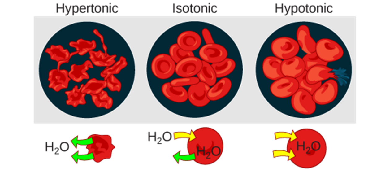
- Hypotonic –
- Hypertonic –
- Isotonic –
For the figure below label each cell correctly hypertonic, hypotonic or isotonic.
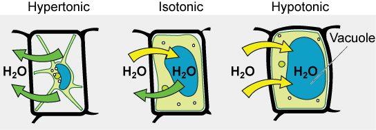
__________________ ________________ __________________
3.6 Active Transport
- How do active transport mechanisms differ from passive transport?
- Briefly explain what is meant by an electrochemical gradient.
- What is primary active transport? List an example as it occurs in cells.
- What is secondary active transport? List an example that occurs in cells.
- Exocytosis –
- Endocytosis –
- Phagocytosis –
- Pinocytosis –
- Receptor-mediated endocytosis –
Make flashcards of the key terms listed at the back of this chapter