The trachea bifurcates at the levels of the 4th-6th intercostal space, approximately …
The trachea bifurcates at the levels of the 4th-6th intercostal space, approximately halfway between the thoracic inlet and the diaphragm. It divides into two principle bronchi, tubes which conduct air into the lungs, and they divide into two lobar bronchi for the left lung, and into four lobar bronchi for the right lung. These further divide into smaller bronchi and bronchioles within the lung tissue.
The air in the alveoli is renewed regularly, thanks to the ventilation …
The air in the alveoli is renewed regularly, thanks to the ventilation process. Gas exchange in the lungs takes place between the blood in the capillary network surrounding the alveoli, and the air in the alveoli itself.
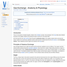
Muscle cells can come from two lineages in the somite. Limb and …
Muscle cells can come from two lineages in the somite. Limb and body muscle develop from hypaxial muscle in the lateral regions of the somite. Back muscle develops from epaxial muscle in the dorsal regions of the somite. Muscle fibres have hundreds of nuclei and function as a syncytium.
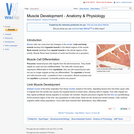
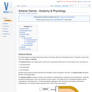
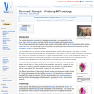
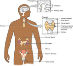
The adrenal glands are paired bodies lying cranial to the kidneys within …
The adrenal glands are paired bodies lying cranial to the kidneys within the retroperitoneal space. The glands consist of two layers; the cortex and medulla.
The adrenal glands are paired bodies lying cranial to the kidneys within …
The adrenal glands are paired bodies lying cranial to the kidneys within the retroperitoneal space. The glands consist of two layers; the cortex and medulla.
The musculoskeletal system includes bones, joints, cartilage, muscles, ligaments and tendons. In …
The musculoskeletal system includes bones, joints, cartilage, muscles, ligaments and tendons. In order to describe anatomical landmarks for example for the purposes of surgery and to be able to describe different directional information, for example when recording the view of a recently taken x-ray, it is necessary to have a way of describing the planes and axes that can be applied to the musculoskeletal system to pinpoint a specific anatomical area.
The ruminant stomach is composed of 4 separate compartments. Food passes first …
The ruminant stomach is composed of 4 separate compartments. Food passes first into the rumen, then reticulum, omasum and finally into the abomasum before entering the duodenum. The first three compartments are adapted to digest complex carbohydrates with the aid of microorganisms which produce volatile fatty acids - the major energy source of ruminants. The last compartments, the abomasum resembles the simple monogastric stomach in structure and function.
These course modules are meant to accompany the OpenStax Anatomy & Physiology …
These course modules are meant to accompany the OpenStax Anatomy & Physiology textbook. Included within each subunit are both Articulate Rise 360 exported raw Web and SCORM 1.2 ZIP files. These files are to be Imported into a Learning Management System. Each module contains text and images from the OpenStax book, original text, openly licensed images from various sources, formative activities, and links to videos on public websites. The modules are free to use as needed. If modification is desired, please contact the author, and I will send you the Rise 360 source file.
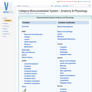
Motor pathways carry signals from the brain to skeletal muscle and smooth …
Motor pathways carry signals from the brain to skeletal muscle and smooth muscle such as those contained in glands. The system consists of upper and lower motor neurones. The information provided below is primarily focussed on the motor pathways that coordinate skeletal muscle movement, i.e. motor pathways related to voluntary control of skeletal muscles.
Corpus Luteum is latin for "yellow body". The corpus luteum is the …
Corpus Luteum is latin for "yellow body". The corpus luteum is the structure formed during luteinisation of the follicle after ovulation. The corpus luteum is, however, actually only yellow in the cow and in all other domestic species it is red. The yellow colouration of the corpus luteum is due to the pigment, lutein.
The phospholipid bilayer is the fundamental structure which makes up the cell …
The phospholipid bilayer is the fundamental structure which makes up the cell membrane. It is made of 2 sheets of phospholipid molecules which are said to have hydrophillic heads and hydrophobic tails. Therefore molecules on opposite sheets face back to back to protect their hydrophobic area from the surrounding intra or extracellular fluid. This creates a region inside the membrane which is hydrophobic.
Idioms that use anatomy vocabulary: Imagine being a new English learner and …
Idioms that use anatomy vocabulary: Imagine being a new English learner and being told to break a leg or pick his brain. Idioms that use anatomy can be weird and downright scary, so this lesson is a must for your ESL students.If you want additional lesson plans and support, including teachers’ notes, be sure to register for a free Off2Class account.
The peritoneum is the serous membrane that lines the abdominal cavity. It …
The peritoneum is the serous membrane that lines the abdominal cavity. It lies directly beneath the abdominal musculature (rectus abdominis and transverse abdominis). It is a type of loose connective tissue and is covered by mesothelium. Extensions of the peritoneum form the mesenteries, omenta and ligaments that support the abdominal contents. The peritoneum produces fluid to lubricate abdominal viscera. The peritoneum also enhances immune responses and walls off infection in the abdomen to prevent peritonitis.
Different hormones, neurotransmitters and reflexes are involved in the complicated process of …
Different hormones, neurotransmitters and reflexes are involved in the complicated process of feeding in animals. Secretions and motility of the gastrointestinal tract are stimulated and carefully regulated by numerous factors, including environmental stimuli and the presence of food in different parts of the gastrointestinal tract from the oral cavity right through to the intestines. When a harmful substance is ingested the body acts to eliminate it in different ways to prevent the animal becoming ill, for example, through vomiting and diarrhoea. If one or more of the pathways in controlling feeding is damaged or inhibited, then problems such as obesity occurs.
After emerging from the heart, the aortic artery divides into the right …
After emerging from the heart, the aortic artery divides into the right and left dorsal branches. Each branch feeds into a set of arches which are unique to the embryo. Most higher vertebrates have have 6 pairs of aortic arches. In the mammal the 5th pair do not form. These arches evolve to form some of the structures of the mammalian circulation. The fate of each arch varies.
The paranasal sinuses are ventilated spaces connected to the nasal cavity. They …
The paranasal sinuses are ventilated spaces connected to the nasal cavity. They develop as blind ending pouches between the lamina of the bones of the skull.
Respiratory epithelium is a type of epithelium which lines both the upper …
Respiratory epithelium is a type of epithelium which lines both the upper and lower respiratory tracts. It consists of multiple layers of cylindrical epithelium, along with cilia and goblet cells.
Development of the Central Nervous System (CNS) includes development of the brain, …
Development of the Central Nervous System (CNS) includes development of the brain, spinal cord, optic and auditory systems, as well as surrounding supporting cells including ependymal cells, astrocytes, oligodendrocytes and microglia. Information within this page will exclude development of the Peripheral Nervous System (PNS) which includes nerve and ganglia formation.
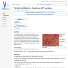
The parathyroid glands are multiple (generally four) small glands, approximately 1-2mm in …
The parathyroid glands are multiple (generally four) small glands, approximately 1-2mm in length are located about the cranial trachea. Generally, there are two internal glands embedded within the thyroid Glands, and two external glands are outside the thyroid tissue. However, all of the parathyroid tissue may be embedded within the thyroid gland itself. In the horse, there are 'nests' of parathyroid tissue along the neck to the thoracic inlet.
No restrictions on your remixing, redistributing, or making derivative works. Give credit to the author, as required.
Your remixing, redistributing, or making derivatives works comes with some restrictions, including how it is shared.
Your redistributing comes with some restrictions. Do not remix or make derivative works.
Most restrictive license type. Prohibits most uses, sharing, and any changes.
Copyrighted materials, available under Fair Use and the TEACH Act for US-based educators, or other custom arrangements. Go to the resource provider to see their individual restrictions.