The Peripheral Nervous System is made up of cranial and spinal nerves. …
The Peripheral Nervous System is made up of cranial and spinal nerves. Spinal nerves are named after the vertebra immediately above it, except for cervical vertebra. There are 7 cervical vertebrae and 8 cervical spinal nerves. The peripheral nervous system can be divided into the somatic nervous system and autonomic nervous system.
A reflex arc represents a mechanism by which a physiological function is …
A reflex arc represents a mechanism by which a physiological function is automatically managed or regulated. Reflex arcs can be found throughout the body, ranging from skeletal muscles to smooth muscle in glands. Reflex arcs are initiated via the excitation or stimulation of specific sensory cells that are directly connected to motor neurons thus enabling motor nerve impulses to be automatically passed on to that particular muscle or gland. Therefore a basic reflex arc consists of sensory cells and their associated nerve fibers, motor nerve fibres and the ultimate muscle or gland.
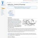
The vagina constitutes the part of the female reproductive tract between the …
The vagina constitutes the part of the female reproductive tract between the cervix and the vulva. With the vestibule and vulva, it is the copulatory organ and the birth canal. The hymen is the poorly developed, vestigial, mucosal folds at the junction of the vagina and vestibule.
The horse is a monogastric hindgut fermenter. The horse evolved for grazing …
The horse is a monogastric hindgut fermenter. The horse evolved for grazing and it does so for up to 17 hours a day. A high proportion of the horse's dietary carbohydrate is in the form of starch. A mature horse eats 2-2.5% of it's body weight in dry matter every day, 1.5-1.75% of this should be fibre (hay/haylage). This is to prevent a rapid drop in pH in the large intestine and also to stimulate peristalsis in the gut and prevent build up of gas.
Word Count: 32276 (Note: This resource's metadata has been created automatically by …
Word Count: 32276
(Note: This resource's metadata has been created automatically by reformatting and/or combining the information that the author initially provided as part of a bulk import process.)
The peritoneum is the serous membrane that lines the abdominal cavity. It …
The peritoneum is the serous membrane that lines the abdominal cavity. It lies directly beneath the abdominal musculature (rectus abdominis and transverse abdominis). It is a type of loose connective tissue and is covered by mesothelium. Extensions of the peritoneum form the mesenteries, omenta and ligaments that support the abdominal contents. The peritoneum produces fluid to lubricate abdominal viscera. The peritoneum also enhances immune responses and walls off infection in the abdomen to prevent peritonitis.
Different hormones, neurotransmitters and reflexes are involved in the complicated process of …
Different hormones, neurotransmitters and reflexes are involved in the complicated process of feeding in animals. Secretions and motility of the gastrointestinal tract are stimulated and carefully regulated by numerous factors, including environmental stimuli and the presence of food in different parts of the gastrointestinal tract from the oral cavity right through to the intestines. When a harmful substance is ingested the body acts to eliminate it in different ways to prevent the animal becoming ill, for example, through vomiting and diarrhoea. If one or more of the pathways in controlling feeding is damaged or inhibited, then problems such as obesity occurs.
After emerging from the heart, the aortic artery divides into the right …
After emerging from the heart, the aortic artery divides into the right and left dorsal branches. Each branch feeds into a set of arches which are unique to the embryo. Most higher vertebrates have have 6 pairs of aortic arches. In the mammal the 5th pair do not form. These arches evolve to form some of the structures of the mammalian circulation. The fate of each arch varies.
The paranasal sinuses are ventilated spaces connected to the nasal cavity. They …
The paranasal sinuses are ventilated spaces connected to the nasal cavity. They develop as blind ending pouches between the lamina of the bones of the skull.
Respiratory epithelium is a type of epithelium which lines both the upper …
Respiratory epithelium is a type of epithelium which lines both the upper and lower respiratory tracts. It consists of multiple layers of cylindrical epithelium, along with cilia and goblet cells.
Development of the Central Nervous System (CNS) includes development of the brain, …
Development of the Central Nervous System (CNS) includes development of the brain, spinal cord, optic and auditory systems, as well as surrounding supporting cells including ependymal cells, astrocytes, oligodendrocytes and microglia. Information within this page will exclude development of the Peripheral Nervous System (PNS) which includes nerve and ganglia formation.
The parathyroid glands are multiple (generally four) small glands, approximately 1-2mm in …
The parathyroid glands are multiple (generally four) small glands, approximately 1-2mm in length are located about the cranial trachea. Generally, there are two internal glands embedded within the thyroid Glands, and two external glands are outside the thyroid tissue. However, all of the parathyroid tissue may be embedded within the thyroid gland itself. In the horse, there are 'nests' of parathyroid tissue along the neck to the thoracic inlet.
Sensory information from the periphery of the animal ascends through the spinal …
Sensory information from the periphery of the animal ascends through the spinal cord and enters the higher levels of the brain. There are numerous pathways which allow different types of information to be passed to the brain. Types of general somatic sensation include pain, touch, temperature and kinaesthesia (conscious proprioception). This sensory information is sent to one of two destinations; the cerebral cortex or the cerebellum.
Blood is supplied to the brain from a ventral arterial supply in …
Blood is supplied to the brain from a ventral arterial supply in all species; from a circle of arteries called the Circle of Willis (also called the cerebral arterial circle or arterial circle of Willis) which lies ventrally to the hypothalamus where it forms a loose ring around the infundibular stalk. Although the appearance of the circle of Willis is fairly constant amongst mammals, the sources of blood supply to the circle and the direction of flow around the circle are very species specific. Blood is supplied to the brain by the internal carotid artery in dogs and horses whilst in other domestic species the main blood supply is from branches of the maxillary artery.
The mammary gland is a modified sweat gland that nourishes the young. …
The mammary gland is a modified sweat gland that nourishes the young. It consists of the mamma and the teat. Undeveloped in both the male and female at birth, the female mammary gland begins to develop as a secondary sex characteristic at puberty.
Osmosis is the passive movement of water across a semi permeable membrane. …
Osmosis is the passive movement of water across a semi permeable membrane. It occurs in the opposite direction to diffusion of ions. Water moves from a region of low solute concentration and therefore high water concentration to a region of high solute concentration and low water concentration.
The formation of the mammalian heart is a fairly complex process. It …
The formation of the mammalian heart is a fairly complex process. It begins when angiogenic mesodermal cells in the cardiogenic plate coalesce to form the endocardial tubes. The endocardial tubes then fuse to form a single duct, the cardiac tube. This undergoes a process of distension, folding and septation and a four chambered, dual circuit pump is formed . The simple heart seen in fish or amphibians forms via the same path but development ceases at an earlier stage.
The hindlimb deep veins are very closely related to their respective arteries. …
The hindlimb deep veins are very closely related to their respective arteries. Essentially the lay out of the veins is similar in all domestic species.
No restrictions on your remixing, redistributing, or making derivative works. Give credit to the author, as required.
Your remixing, redistributing, or making derivatives works comes with some restrictions, including how it is shared.
Your redistributing comes with some restrictions. Do not remix or make derivative works.
Most restrictive license type. Prohibits most uses, sharing, and any changes.
Copyrighted materials, available under Fair Use and the TEACH Act for US-based educators, or other custom arrangements. Go to the resource provider to see their individual restrictions.