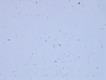
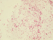
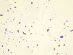
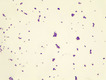
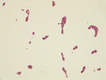
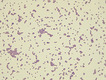
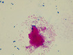
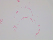
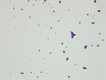
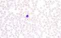
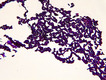
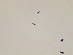
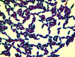
All resources in College of Marin

Micrograph Escherichia coli and Mycobacterium smegmatis acid fast 1000X p000173

(View Complete Item Description)

This micrograph was taken at 1000X total magnifcation on a brightfield microscope. The subject is Neisseria sicca cells and Mycobacterium phlei cells grown separately in broth culture at 37 degrees Celsius. The cells were heat-fixed to a slide and acid-fast stained using the Ziehl-Neelsen method prior to visualization. M. smegmatis (acid-fast) stains pink with carbol fuchsin. N. sicca (non-acid-fast) stains blue with methylene blue counterstain.Image credit: Emily Fox

Material Type: Diagram/Illustration

Author: Emily Fox

Blood Smear Video v000001

(View Complete Item Description)

This video shows scanning a Wright's stained blood smear slide with pauses to view leukocytes. The video was taken at 630X under a brightfield microscope. This video is compatible with a laboratory lesson in which students observe, categorize, and count leukocytes. More than 100 leukocytes are viewed in this video. Note, this video does not have narration.Video credit: Emily Fox

Material Type: Diagram/Illustration

Author: Emily Fox