
Unrestricted Use
CC BY
OpenStax Anatomy & Physiology Quizlet list
- Subject:
- Anatomy/Physiology
- Material Type:
- Diagram/Illustration
- Author:
- Rob Adams
- Date Added:
- 05/05/2021
OpenStax Anatomy & Physiology Quizlet list

These are interactive activities that are based on the OpenStax textbook on Anatomy and Physiology. The majority of definitions have been incorporated into dialogue cards. And the majority of diagrams have been made into interative drag and drop with MCQ and other question types.

Brehe’s Grammar Anatomy makes grammar accessible to general and specialist readers alike. This book provides an in-depth look at beginner grammar terms and concepts, providing clear examples with limited technical jargon. Whether for academic or personal use, Brehe’s Grammar Anatomy is the perfect addition to any resource library.

This zipped folder contains 28 Power Point files that correspond to each of the chapters in the OpenStax Anatomy and Physiology textbook. These are meant to provide a starting point for presentation files related to an Anatomy and Physiology course. The design should be easily modified using the “design” tab in Microsoft Power Point. The end user should be able to quickly choose a template/color scheme that works for them. Additionally, the end user may want to add or remove text from each power point slide. This can easily be accomplished by simply editing the document. Most of the images used in the creation of these Power Points are taken directly from the OpenStax Anatomy and Physiology textbook. Supplemental images pulled from elsewhere include a small textbox with a link to the original work and the CC license terms.

The overall purpose of this preparatory course textbook is to help students familiarize with some terms and some basic concepts they will find later in the Human Anatomy and Physiology I course.
The organization and functioning of the human organism generally is discussed in terms of different levels of increasing complexity, from the smallest building blocks to the entire body. This Anatomy and Physiology preparatory course covers the foundations on the chemical level, and a basic introduction to cellular level, organ level, and organ system levels. There is also an introduction to homeostasis at the beginning.
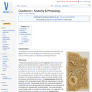
The duodenum is the proximal part of the small intestine and extends from the pylorus of the stomach to the jejunum. It has descending and ascending portions and both portions have digestive and absorptive functions.
The jejunum continues from the duodenum and leads into the ileum. It is the longest part of the small intestine and is highly coiled. It has digestive and absorptive functions.
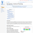
Nerves allow electrical impulses to propagate along their elongated cell extensions and facilitate the transfer of information throughout the body. Neural tissue is found within the central nervous system (CNS) and the peripheral nervous system (PNS) and the composition and constituent parts of neurones and their surrounding cells differ only slightly.
The keratin in the epidermis, when cornified and thickened, is referred to as horn. Horn is particulary resistant to mechanical and chemical damage. The dermis of horn gives the structures their 3-D structure and shape. Cattle, some sheep, goats and antelope posess horns and these are permanent organs. Breeds without horns are termed polled breeds. Deer posess antlers, which are temporary organs that develop during the rutting season and are then shed.
The ovary is the female Gonad homologous to the male Testes. It is usually a paired organ in domestic species, but in the bird only the left Ovary is present. The structures found within the ovary are undergoing constant changes throughout the oestrus cycle from the Follicles containing Oocytes, to the formation of Corpus Haemorrhagicum, Corpus Luteum, and finally Corpus Albicans. Ovaries are ellipsoidal in shape with an irregular surface due to the projection of dominant follicles and corpora lutea. These irregularities are absent in the mare due to the cortex and medulla being reversed with ovulation only occuring from the ovulation fossa. They are greatest in Polytocious animals such as the sow due to many dominant follicles, and so corpora lutea, developing at once.
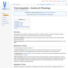
Thermoregulation is the ability of an endothermic organism to maintain a relatively constant body temperature, despite fluctuations in temperature of the external environment. This is a vital part of homeostasis.
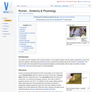
The rumen is the first chamber of the ruminant stomach. It is the largest chamber and has regular contractions to move food around for digestion, eliminate gases through eructation and send food particles back to the mouth for remastication. The rumen breaks down food particles through mechanical digestion and fermentation with the help of symbiotic microbes. Volatile fatty acids are the main product of ruminant digestion.
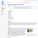
The omasum is the third chamber in the ruminant stomach. It lies within the intrathoracic part of the abdomen so cannot be palpated manually. Instead it is examined by ausculation. The omasum has biphasic contractions which squeeze fluid out of the food before allowing the ingesta to continue into the abomasum. Absorption of volatile fatty acids continues in the omasum.
The rectum lies between the terminal portion of the descending colon and anus. It is empty most of the time, except after the mass movements of the large intestine which move faeces into the rectum. This stimulates defeaction, which may happen when an animal is frightened.
The liver (hepar) is an extremely important organ in the body of mammals and vertebrates as it provides functions essential for life. It is the largest internal organ and has numerous functions including production of bile and protein, fat and carbohydrate metabolism. During foetal development, the liver has an important haemopoetic function, producing red and white blood cells from tissue between the hepatic cells and vessel walls.
The term 'implantation' is often used to describe the attachment of the placental membranes to the endometrium in most animals. True implantation is a phenomenon in rodents and humans in which the conceptus 'buries' itself in the uterine endometrium. The conceptus temporarily disappears beneath the surface. In most other species, the conceptus does not truly implant, but attaches to the endometrial surface and remains in the luminal compartment.
Hair germs begin from an aggregation of keratinocytes in the stratum basale of the epidermis. The initiating factor is the underlying dermal fibroblast cells. The keratinocytes elongate, divide and relocate to the dermis. Dermal fibroblasts then form a dermal papilla beneath the hair germ. This causes stimulation of the basal stem cells to up-regulate their cycle, producing cells that will keratinise and form the hair shaft.
The epidermis is a stratified squamous epithelium and is composed of 4 cell layers anchored to a basal lamina of connective tissue. Keratinocytes migrate through the epidermis from the basal layer. This migration begins in the stratum basale, then moves up through the stratum spinosum, stratum granulosum and the stratum corneum.
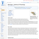
The central nervous system (CNS) is surrounded by several layers of tissue, with several outer layers not directly related to the CNS and three membranes that directly envelope the CNS. The outer layers are the skin and then a bone layer with associated periosteum. This layer includes the skull and the vertebrae. Below the periosteum is the Epidural Space which lies between periosteum and dura in the vertebral canal. The epidural space contains adipose tissue, loose connective tissue, veins and lymphatics. It cushions the cord as it flexes.
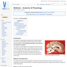
The midbrain or mesencephalon represents the connection between the brain stem and the higher centres of the brain and is involved in most body systems including sleep/consciousness, vision, hearing and temperature regulation.