(This case study was added to OER Commons as one of a …
(This case study was added to OER Commons as one of a batch of over 700. It has relevant information which may include medical imagery, lab results, and history where relevant. A link to the final diagnosis can be found at the end of the case study for review. The first paragraph of the case study -- typically, but not always the clinical presentation -- is provided below.)
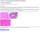
A 29-year-old man described progressive difficulty climbing stairs over a decade. He did not report any other neurological symptoms including upper limb, bulbar, facial or respiratory muscle weakness. His perinatal history and development were unremarkable, although he had never been particularly good at sport. There was no family history of cardiac or neuromuscular disease. Neurological examination revealed an exaggerated lumbar lordosis and myopathic gait. He had moderate symmetrical, predominantly proximal, limb weakness which was more marked in the lower than the upper limbs and normal tendon reflexes. There was no sensory loss, myotonia or muscle hypertrophy. Serum creatine kinase level was elevated at 1096 IU/L (upper limit of normal: 250 IU/L). Electromyogram was myopathic. Electrocardiogram and echocardiography were normal.
(This case study was added to OER Commons as one of a …
(This case study was added to OER Commons as one of a batch of over 700. It has relevant information which may include medical imagery, lab results, and history where relevant. A link to the final diagnosis can be found at the end of the case study for review. The first paragraph of the case study -- typically, but not always the clinical presentation -- is provided below.)
The patient is a previously healthy 29-year-old man who was admitted to our hospital for evaluation after experiencing seizures. A neurological examination elicited no abnormalities. CT and MR imaging were performed and revealed a cortico-subcortical lesion mass involving most of the right frontal lobe with sulcal effacement, compression on the foramen of Monro and hydrocephalus. The CT demonstrated curvilinear narrow calcifications (Figure 1). The lesion was hyperintense to the cortex on T2 and FLAIR (Figures 2 and 3) while partially hyperintense on T1 (Figure 4), with a marked homogeneous enhancement (Figure 5). All the sequences showed a "bag-of-worms sign" (which is best seen on the T2-weighted images) due to the presence of multiple tiny vessels associated with flow-voids (Figures 2 and 3). Moreover, in the lesion there were two deeply seated pseudocysts that showed contrast enhancement visible in the late phase. Based on these findings, a digital subtraction angiography (DSA) was performed which confirmed the hypervascular nature of the lesion but it did not indicate the presence of an arteriovenous malformation (Figure 6). At surgery, the lesion was removed via a right frontal craniotomy. The excision was macroscopically complete.
(This case study was added to OER Commons as one of a …
(This case study was added to OER Commons as one of a batch of over 700. It has relevant information which may include medical imagery, lab results, and history where relevant. A link to the final diagnosis can be found at the end of the case study for review. The first paragraph of the case study -- typically, but not always the clinical presentation -- is provided below.)
A 26-year-old man presented with a 1 year history of memory impairment. He became somnolence and lethargy 8 months before seeking medical attention. He needed to sleep over 20 hours per day. The physical examination and neurological examination were unremarkable.
(This case study was added to OER Commons as one of a …
(This case study was added to OER Commons as one of a batch of over 700. It has relevant information which may include medical imagery, lab results, and history where relevant. A link to the final diagnosis can be found at the end of the case study for review. The first paragraph of the case study -- typically, but not always the clinical presentation -- is provided below.)
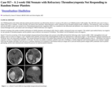
The patient was a 29 year old Caucasian female with a past medical history of poorly controlled hypertension and diabetes mellitus who presented with a one week history of marked confusion and ataxia. She was brought to the hospital by local authorities who found her wandering in a nearby creek, completely disoriented with regards to where she was or how she had gotten there.
(This case study was added to OER Commons as one of a …
(This case study was added to OER Commons as one of a batch of over 700. It has relevant information which may include medical imagery, lab results, and history where relevant. A link to the final diagnosis can be found at the end of the case study for review. The first paragraph of the case study -- typically, but not always the clinical presentation -- is provided below.)
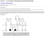
A 2-month-old male infant was referred to our hospital due to congenital hypotonia. The patient was normally delivered at 42 weeks of gestation age to a 20-year-old healthy mother. He was the first child of this family. Based on regular antenatal care, no abnormal findings were reported until birth when the APGAR scores at 1 and 5 minutes were 4 and 7, respectively. He had 2,850 g birthweight, 49 cm crown heel length, and 34 cm head circumference. The patient was hypotonic and had respiratory distress which required endotracheal intubation soon after delivery. Physical examination revealed generalized hypotonia. Myopathic facies without high-arched palate was noted. There was no evidence of metabolic disorders. Serum creatine kinase level was normal (56 IU/L). Serology tests for congenital infections were negative. SMN gene analysis displayed no mutation suggestive of spinal muscular atrophy. There was no evidence of consanguineous marriage in his family. The mother and her sister are clinically normal. The mother's male siblings passed away at 1 month and 1.5 months after birth due to respiratory distress complicated by respiratory infection (Figure 1).
(This case study was added to OER Commons as one of a …
(This case study was added to OER Commons as one of a batch of over 700. It has relevant information which may include medical imagery, lab results, and history where relevant. A link to the final diagnosis can be found at the end of the case study for review. The first paragraph of the case study -- typically, but not always the clinical presentation -- is provided below.)
A 2-year-old girl presented with one-month history of dysmetria and ataxia. Computer tomography (CT) of the brain showed a large parietal-occipital mass and marked hydrocephalus, for which she required placement of bilateral ventriculoperitoneal shunts. Magnetic resonance imaging (MRI) of the brain also revealed two nodules within the lateral ventricle (Figs. 1, 2 and 3, axial T1, non-enhanced T2, diffusion weighted imaging, respectively). Spine MRI showed abnormal leptomeningeal enhancement along the cervical spine, with additional enhancement in the thoracic and upper lumbar spine. A metastatic origin was excluded by exhaustive radiologic investigations, including body CT and positron emission tomography. Her disease progressed following completion of 6 months of intravenous and 3 months of oral chemotherapy; MRI showed interval increase in the size of the left temporal lobe ventricular lesions as well as worsening leptomeningeal disease and new parenchymal nodules. The tumor was completely excised and submitted for histopathological examination.
(This case study was added to OER Commons as one of a …
(This case study was added to OER Commons as one of a batch of over 700. It has relevant information which may include medical imagery, lab results, and history where relevant. A link to the final diagnosis can be found at the end of the case study for review. The first paragraph of the case study -- typically, but not always the clinical presentation -- is provided below.)
An O Rh(D)-positive term infant with intra-uterine growth restriction was delivered by elective C-section at 38 weeks to an O Rh(D)-positive G2P2 mother. The APGAR scores were 9 /10 at 1 minute and 9 /10 at 5 minutes. At birth, the baby was noted to have petechiae. Initial laboratory results confirmed severe thrombocytopenia (nadir 10,000/µL). The hematocrit, white cell (WBC) and red cell (RBC) counts were normal to low. Disseminated intravascular coagulation (DIC) was considered in the differential and antibiotics were initiated; however, the DIC workup showed normal fibrinogen levels, and cultures and viral testing for TORCH infections were negative.
(This case study was added to OER Commons as one of a …
(This case study was added to OER Commons as one of a batch of over 700. It has relevant information which may include medical imagery, lab results, and history where relevant. A link to the final diagnosis can be found at the end of the case study for review. The first paragraph of the case study -- typically, but not always the clinical presentation -- is provided below.)
A 2-year-old boy presented with gait imbalance and strabismus over the last two weeks. Five days before admission he began to vomit and became irritable. On admission a brain computed tomography was performed and revealed a large midline posterior fossa mass; the lesion was heterogeneous, cystic partly solid with calcifications (Figure 1). Magnetic resonance imaging that ensued demonstrated a 5x4.9x4.7cm mass within the 4th ventricle, extending through foramen of Luschka into the left cerebello-pontine angle without infiltration of the vermis (Figure 2). The lesion showed heterogenous enhancement after contrast administration (Figure 3). The patient was operated upon and a gross total resection of the mass was performed.
(This case study was added to OER Commons as one of a …
(This case study was added to OER Commons as one of a batch of over 700. It has relevant information which may include medical imagery, lab results, and history where relevant. A link to the final diagnosis can be found at the end of the case study for review. The first paragraph of the case study -- typically, but not always the clinical presentation -- is provided below.)
A previously healthy 2-year-old boy presented with one to two days of anuria and bloody diarrhea. He was admitted to the local children's hospital with a diagnosis of hemolytic uremic syndrome (HUS), presumably secondary to E. coli O157 but never confirmed. Laboratory studies revealed hemolysis, renal failure and thrombocytopenia. Eleven hours after admission, his blood oxygen saturations worsened and he was intubated. He was then noted to have fixed and dilated pupils. A head CT was performed, which revealed left frontal subcortical white matter vasogenic edema with left frontal gyral hyperdensity, as well as scattered pockets of pneumocephalus (Figure 1). Pneumocephalus without evidence of a calvarial fracture raised concern for an infectious process. Left-to-right midline shift with effacement of the cisterns and left uncal herniation were also observed. Neurosurgical consultation advised that no intervention was possible. The patient became bradycardic and required resuscitation, which unfortunately was not successful. He expired 14 hours after being admitted to the hospital. Ante-mortem bacterial blood cultures were positive.
(This case study was added to OER Commons as one of a …
(This case study was added to OER Commons as one of a batch of over 700. It has relevant information which may include medical imagery, lab results, and history where relevant. A link to the final diagnosis can be found at the end of the case study for review. The first paragraph of the case study -- typically, but not always the clinical presentation -- is provided below.)
This 2-year-old male was referred to a Genetic Counselor at the age of 22 months for developmental delay and dysmorphic features.
(This case study was added to OER Commons as one of a …
(This case study was added to OER Commons as one of a batch of over 700. It has relevant information which may include medical imagery, lab results, and history where relevant. A link to the final diagnosis can be found at the end of the case study for review. The first paragraph of the case study -- typically, but not always the clinical presentation -- is provided below.)
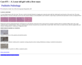
A 2-year old girl presents with a liver mass. The mass is unifocal and multi-lobulated on imaging and occupies the entire right lobe, displacing the main branches of the portal veins. The serum alpha-fetoprotein is elevated to 680 ng/dL and the only other pertinent finding is an elevated platelet count to 685,000. An open biopsy is performed and representative sections are displayed below:
(This case study was added to OER Commons as one of a …
(This case study was added to OER Commons as one of a batch of over 700. It has relevant information which may include medical imagery, lab results, and history where relevant. A link to the final diagnosis can be found at the end of the case study for review. The first paragraph of the case study -- typically, but not always the clinical presentation -- is provided below.)
A 30-year-old female has experienced amenorrhea and progressive loss of vision for four years. Physical examinations were normal except bitemporal hemianopsia revealed by ophthalmic examination. Preoperative neuroendocrine examinations showed a mild hyperprolactinemia of 72.3ng/ml (normal range, 2.8 ng/ml-29.2 ng/ml). MRI scan revealed a 31 mm × 34 mm × 31 mm well-circumscribed roundness mass in the suprasellar region, with intermediate signal intensity on T1-weighted images, intermediate to slightly increased signal intensity on T2-weighted images, and homogeneous enhancement with gadolinium administration with obviously homogeneous enhancement after gadolinium administration (Fig. 1). Extended endoscopic endonasal transsphenoidal approach was chosen to resect the tumor. Intraoperatively, we encountered active bleeding, however, the bleeding stopped after the tumor was completely resected. Postoperative, the patient had serious diabetes insipidus and electrolyte disturbance. Blood sodium was as high as 190mmol/l (normal range, 135 mmol/L-145 mmol/L). After comprehensive treatment, the diabetes insipidus was cured and blood sodium became normal. Three months after endoscopic surgery, MRI examination revealed that the tumor was completely removed (Fig. 2).
(This case study was added to OER Commons as one of a …
(This case study was added to OER Commons as one of a batch of over 700. It has relevant information which may include medical imagery, lab results, and history where relevant. A link to the final diagnosis can be found at the end of the case study for review. The first paragraph of the case study -- typically, but not always the clinical presentation -- is provided below.)
A 30 year-old man presented with progressive visual field defect, headache and nausea. He had a history of occipital area blunt head injury (hit by an iron pipe at work) three months before referral to our clinic. The initial diagnosis at the local clinic was brain contusion and concussion. On the initial X ray no skull fracture was detected. However, owing to a progressive visual field defect and dizziness, epidural hemorrhage was suspected. A burr-hole trephination for drainage was subsequently performed. Unexpectedly, the operating neurosurgeon found a tumor mass instead of hematoma. Although the biopsy sample was obtained, only H&E slides were made without ancillary immunohistochemical stains (IHC). He was then transferred to our hospital for further evaluation and more tissue was obtained. The patient had no history of malignancy. All laboratory findings were within normal ranges, except AST 67 U/L (Normal range: 14-40), ALT 164 U/L (Normal range: 8-46) and serum lactated dehydrogenase (LDH) 532 U/L (Normal range: 218-472). Serologic tests for hepatitis, syphilis, and HIV infection were negative. Magnetic resonance imaging (MRI) revealed an epidural mass, approximately 7.0x6.8x4.1 cm, with extracranial extension forming a coffee bean shaped mass crossing the parieto-occipital lobe dura (Figs. 1, 2 and 3). The epidural mass was hyperintense to isointense to gray matter on T2 weighted images (WI) (Fig. 1), isointense on T1WI (Fig. 2). Following contrast infusion, the epidural mass showed marked heterogeneous enhancement but the extracranial mass did not (Fig. 3). In view of the radiologic findings, a clinical diagnosis of meningioma was considered and the patient underwent a brain biopsy to confirm the nature of the brain lesion.
(This case study was added to OER Commons as one of a …
(This case study was added to OER Commons as one of a batch of over 700. It has relevant information which may include medical imagery, lab results, and history where relevant. A link to the final diagnosis can be found at the end of the case study for review. The first paragraph of the case study -- typically, but not always the clinical presentation -- is provided below.)
The patient is a 30 year-old female with history of familial polyposis, status post transanal excision of rectal villous adenoma, and restorative proctocolectomy with ileoanal anastomosis and loop ileostomy for colonic polyposis. An upper gastrointestinal endoscopy was performed and demonstrated multiple gastric polyps in the fundus and body (Figure 1A and B). Representative biopsies from the gastric polyps were submitted for pathological examination.
(This case study was added to OER Commons as one of a …
(This case study was added to OER Commons as one of a batch of over 700. It has relevant information which may include medical imagery, lab results, and history where relevant. A link to the final diagnosis can be found at the end of the case study for review. The first paragraph of the case study -- typically, but not always the clinical presentation -- is provided below.)
The patient is a 30-year-old male with a 2-month history of a right corneal abrasion when removing his contact lens. Initial bacterial and fungal culture results were negative, and when he returned for a follow-up appointment, he complained of increasing ocular pain, headaches, and vision loss. Physical exam revealed an abrasion that had failed to heal, leading to a purulent corneal ulcer. Ocular fluid was collected, and the patient was started on antibiotics for a suspected bacterial infection.
(This case study was added to OER Commons as one of a …
(This case study was added to OER Commons as one of a batch of over 700. It has relevant information which may include medical imagery, lab results, and history where relevant. A link to the final diagnosis can be found at the end of the case study for review. The first paragraph of the case study -- typically, but not always the clinical presentation -- is provided below.)
A 30-year-old Chinese male presented with an 8 week history of headaches, visual changes, lightheadedness and balance problems. These symptoms were attributed to increased intracranial pressure secondary to obstructive hydrocephalus. Computed tomography (CT) scans demonstrated an iso-or slightly hyperdense mass within both lateral ventricles. The septum pellucidum was not seen well on imaging studies and the tumor was centered on the midline. Contrast enhancement was mild to moderate. An MRI scan revealed a large midline mass obliterating large portions of the lateral ventricles and giving rise to obstructive hydrocephalus and transependymal edema. The mass was isointense on T1 weighted imaging and contained calcification On T2 weighted images it was relatively isointense with cortex (Fig. 1 and Fig. 2). There was moderate enhancement after the administration of gadolinium. Postoperative MRI scan showed gross total resection and some blood. After radiotherapy, the nerval symptoms had lapse to. But repeated MRI scans in the following 12 months showed recrudescence in the MRI findings and patient had more nerval symptomatic. Finally, the patient died of hydrocephalus and brain edema 20 months after the surgery.
(This case study was added to OER Commons as one of a …
(This case study was added to OER Commons as one of a batch of over 700. It has relevant information which may include medical imagery, lab results, and history where relevant. A link to the final diagnosis can be found at the end of the case study for review. The first paragraph of the case study -- typically, but not always the clinical presentation -- is provided below.)
The patient is a 30-year old man with a 30-pound weight loss in one month, poor appetite, and episodes of abdominal and back pain. He has had progressive pancytopenia over 1 month.
(This case study was added to OER Commons as one of a …
(This case study was added to OER Commons as one of a batch of over 700. It has relevant information which may include medical imagery, lab results, and history where relevant. A link to the final diagnosis can be found at the end of the case study for review. The first paragraph of the case study -- typically, but not always the clinical presentation -- is provided below.)
A 30-years old male patient had suffered from childhood epilepsy at the age of three years. He presented with multiple cutaneous angiofibromas, bilateral renal angiomyolipomas and a tumor of the liver that had not been biopsied. His family history was unremarkable; in particular, TSC history was negative.
(This case study was added to OER Commons as one of a …
(This case study was added to OER Commons as one of a batch of over 700. It has relevant information which may include medical imagery, lab results, and history where relevant. A link to the final diagnosis can be found at the end of the case study for review. The first paragraph of the case study -- typically, but not always the clinical presentation -- is provided below.)
A 30-year-old woman presented to her GP with complaints of intermittently occurring joint pain in her wrists and ankles. Further questioning revealed that she had been experiencing occasional morning stiffness and a tingling sensation in her extremities. She denied any headache or constitutional symptoms beyond mild fatigue. She stated that she had been tested for ANA and rheumatoid factor in the past and they were both negative. Additional inquiry revealed that the patient has a positive family history for rheumatoid arthritis. On physical exam, the patient expressed mild tenderness at both wrists. The rest of the exam was non-contributory. The GP was concerned about a potential autoimmune disease process and ordered an erythrocyte sedimentation rate (ESR) and an antinuclear antibody (ANA) test. The results are listed in table 1. The GP then referred the patient to rheumatology.
No restrictions on your remixing, redistributing, or making derivative works. Give credit to the author, as required.
Your remixing, redistributing, or making derivatives works comes with some restrictions, including how it is shared.
Your redistributing comes with some restrictions. Do not remix or make derivative works.
Most restrictive license type. Prohibits most uses, sharing, and any changes.
Copyrighted materials, available under Fair Use and the TEACH Act for US-based educators, or other custom arrangements. Go to the resource provider to see their individual restrictions.