(This case study was added to OER Commons as one of a …
(This case study was added to OER Commons as one of a batch of over 700. It has relevant information which may include medical imagery, lab results, and history where relevant. A link to the final diagnosis can be found at the end of the case study for review. The first paragraph of the case study -- typically, but not always the clinical presentation -- is provided below.)
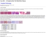
The patient is a female in her 70s with pelvic masses and ascites. Her preoperative CA-125 increased to 130 units/ml, but CA19-9 and CEA were normal. She underwent total abdominal hysterectomy, bilateral salphingo-oophorectomy, omentectomy as well as cul-de-sac excisional biopsy.
(This case study was added to OER Commons as one of a …
(This case study was added to OER Commons as one of a batch of over 700. It has relevant information which may include medical imagery, lab results, and history where relevant. A link to the final diagnosis can be found at the end of the case study for review. The first paragraph of the case study -- typically, but not always the clinical presentation -- is provided below.)
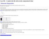
A male in his 40s with a prior medical history of Behcet's disease and severely compromised vision presented with a cognitive disorder and chorea. His symptoms began 8 years ago, over which time the patient has steadily deteriorated. His mother had similar symptoms and was diagnosed as having Huntington disease. Few family members on the maternal side were also affected (see Pedigree). None of the mother's relatives were previously tested.
(This case study was added to OER Commons as one of a …
(This case study was added to OER Commons as one of a batch of over 700. It has relevant information which may include medical imagery, lab results, and history where relevant. A link to the final diagnosis can be found at the end of the case study for review. The first paragraph of the case study -- typically, but not always the clinical presentation -- is provided below.)
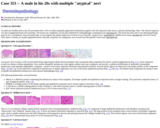
An otherwise healthy male in his 20s presented with a 0.95 cm shiny unequally pigmented erythematous papule in the left supraclavicular area to a private dermatology clinic. The clinical suspicion was that of pigmented basal cell carcinoma. The lesion was completely excised and submitted for histopathologic examination (see specimen #1). The lesion has been sent to our dermatopathology practice for a consultation. Several months later, in our hospital, the patient underwent excision of two clinically "atypical nevi" (specimen #2: midline lower back; specimen #3: left lower back). After about 2 months yet another pigmented lesion clinically suspicious for malignancy was excised from this patient's lower back (specimen #4)
(This case study was added to OER Commons as one of a …
(This case study was added to OER Commons as one of a batch of over 700. It has relevant information which may include medical imagery, lab results, and history where relevant. A link to the final diagnosis can be found at the end of the case study for review. The first paragraph of the case study -- typically, but not always the clinical presentation -- is provided below.)
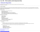
This baby boy with postnatal onset microcephaly had speech impairment and global developmental delay that were noted at 12 months of age. He also had feeding problems which included gagging, choking and frequent drooling. He was born by Cesarean section for breech presentation to a 28-year-old G1P1 subsequent to an unremarkable pregnancy. There is no family history of developmental delay or neurological problems.
(This case study was added to OER Commons as one of a …
(This case study was added to OER Commons as one of a batch of over 700. It has relevant information which may include medical imagery, lab results, and history where relevant. A link to the final diagnosis can be found at the end of the case study for review. The first paragraph of the case study -- typically, but not always the clinical presentation -- is provided below.)
This 42-year-old Caucasian woman was referred for diagnosis and therapy of an ampullary adenoma. The patient had been in good health until 3 months earlier, when she developed episodic right upper quadrant (RUQ) and back pain. On gallbladder ultrasound, there was evidence of calcified gallstones with normal gallbladder wall and bile duct. Laboratory tests revealed a mild elevation in alanine amino-transferase (ALT) (48 U/liter). The CBC, chemistry profile, aspartate aminotransferase (AST), alkaline phosphatase, total bilirubin, lipase, and the urine analysis were normal. Family history includes: mother deceased of breast cancer at age 64, father deceased at age 72 of coronary artery disease. and one sister who died at age 37 of renal cancer (she is one of four children). She has a 14 and a 21-year-old child.
(This case study was added to OER Commons as one of a …
(This case study was added to OER Commons as one of a batch of over 700. It has relevant information which may include medical imagery, lab results, and history where relevant. A link to the final diagnosis can be found at the end of the case study for review. The first paragraph of the case study -- typically, but not always the clinical presentation -- is provided below.)
A 69-year-old man presented with a left neck mass. The patient underwent total thyroidectomy, central compartment neck dissection, sternocleidomastoid muscle flap reconstruction, and primary closure of the esophageal wall. The total thyroidectomy specimen was irregular in shape, the right lobe measured 4.5 x 2.0 x 1.5 cm and the left lobe measures 9.0 x 5.5 x 4.5 cm. The exterior surface of the gland was tan-pink and rubbery. The cut surface of the left lobe revealed a 7.5 x 4.5 cm tan-grey lesion. The lesion approached to within less than 1mm of the capsular surface. Scant normal appearing thyroid parenchyma was present at the superior aspect of the left lobe. The right lobe parenchyma was red and beefy.
(This case study was added to OER Commons as one of a …
(This case study was added to OER Commons as one of a batch of over 700. It has relevant information which may include medical imagery, lab results, and history where relevant. A link to the final diagnosis can be found at the end of the case study for review. The first paragraph of the case study -- typically, but not always the clinical presentation -- is provided below.)
The patient is a 74-year-old Caucasian male, who had intermittent skin rash for seven years. Rash usually goes away with medicated creams, but recurs every year. A skin biopsy was obtained at a local hospital four years ago and was suggestive of mycosis fungoides (cutaneous T-cell lymphoma). He was treated with oral and topical steroids. The topical therapy included prednisone (with a taper), triamcinolone and fluocinonide. In the past 2 years, the patient was found to have anemia (hemoglobin was 10.3 g/dl) with a normal white cell count and thrombocytopenia (96 x 109/L) of unknown etiology. Laboratory studies indicated normal values for reticulocyte count, serum levels of B12, folate, ferritin, and total iron binding capacity. He did have a history of heme-positive stool, but refused colonoscopy. The patient underwent a bone marrow biopsy one and half years ago. The results included a hypercellular marrow with trilineage hematopoiesis, focally increased and dysplastic megakaryocytes, suggestive of myelodysplastic syndrome (MDS) and no increase in blasts. Flow cytometry immunophenotyping did not identify an aberrant T or B cell immunophenotype. Cytogenetic studies were normal. He had been treated with Procrit.
(This case study was added to OER Commons as one of a …
(This case study was added to OER Commons as one of a batch of over 700. It has relevant information which may include medical imagery, lab results, and history where relevant. A link to the final diagnosis can be found at the end of the case study for review. The first paragraph of the case study -- typically, but not always the clinical presentation -- is provided below.)
Our patient is a 68-year-old man who was transferred to this hospital with upper gastrointestinal (GI) bleeding. His medical history was significant for follicular lymphoma involving a left supraclavicular lymph node diagnosed 7 months prior to another lymphoma involving the right chest wall. Lymphoma of the chest wall preceded the upper GI bleeding by 2 months. Treatment for the lymphomas included 6 cycles of CHOP and local radiation to the chest.
(This case study was added to OER Commons as one of a …
(This case study was added to OER Commons as one of a batch of over 700. It has relevant information which may include medical imagery, lab results, and history where relevant. A link to the final diagnosis can be found at the end of the case study for review. The first paragraph of the case study -- typically, but not always the clinical presentation -- is provided below.)
A 19-year-old previously healthy man was admitted in an unconscious state with a conjugate deviation of gaze to the right. Having been intubated and ventilated, he suffered a series of generalized seizures. Cranial MRI showed a slightly enhanced periventricular edema zone in the white matter adjacent to the posterior horn (Fig. 1; A: fluid-attenuated inversion recovery [FLAIR], B: T2-weighed, C: gadolinium enhanced).
(This case study was added to OER Commons as one of a …
(This case study was added to OER Commons as one of a batch of over 700. It has relevant information which may include medical imagery, lab results, and history where relevant. A link to the final diagnosis can be found at the end of the case study for review. The first paragraph of the case study -- typically, but not always the clinical presentation -- is provided below.)
A 61-year-old female with a past medical history of chronic lymphocytic leukemia (CLL) for 10 years, presented to the emergency room (ER) with left upper quadrant pain and multiple ecchymoses. Her past medical history included treatment with Fludarabine (last dose more than one year ago), Rituximab and Cytoxan. Of note, she presented to the ER with a seizure 6 months previously. Studies revealed a left fronto-temporal lesion, which was resected and diagnosed as a meningioma. To prevent seizures, she was started on Dilantin. At a follow up visit in neurosurgery, CBC values indicated pancytopenia. Because this was thought to be related to Dilantin treatment, it was suspended and Levetiracetam was initiated, but there were no improvements in her counts.
(This case study was added to OER Commons as one of a …
(This case study was added to OER Commons as one of a batch of over 700. It has relevant information which may include medical imagery, lab results, and history where relevant. A link to the final diagnosis can be found at the end of the case study for review. The first paragraph of the case study -- typically, but not always the clinical presentation -- is provided below.)
In 1999, at the age of 41 this man developed focal seizures in his right arm. The neurological examination was otherwise normal. He was in good health and his medical history was devoid of underlying disease. Cranial MRI revealed a homogeneously contrast enhancing lesion with microcalcifications in the left frontal lobe (Figs. 1 and 2). One year later the patient decided to have stereotactic biopsy. Surgically induced artifacts and small sample size aggravated tumor classification at that time. Differential diagnoses included oligodendroglioma WHO II and diffuse astrocytoma WHO II. In January 2003, at the age of 45, the patients developed weakness in his right arm and seizure frequency increased despite medication. Tumor size had considerably increased. Surgery was offered and the patient decided to have the lesion removed through a left frontal craniotomy. The postoperative course, was unremarkable, the weakness of the right arm disappeared.
(This case study was added to OER Commons as one of a …
(This case study was added to OER Commons as one of a batch of over 700. It has relevant information which may include medical imagery, lab results, and history where relevant. A link to the final diagnosis can be found at the end of the case study for review. The first paragraph of the case study -- typically, but not always the clinical presentation -- is provided below.)
This is a 54-year-old female with a medical history of emphysema secondary to alpha-1 antitrypsin deficiency, status-post double lung transplant in January 2004. Her post-operative course was complicated by acute cellular rejection (grade 3), severe hemodynamic instability, pneumonia, and a sternal wound infection, requiring multiple, prolonged hospital admissions. During that time she was treated with a variety of immunosuppressants including azathioprine, cyclosporine, tacrolimus, and prednisone.
(This case study was added to OER Commons as one of a …
(This case study was added to OER Commons as one of a batch of over 700. It has relevant information which may include medical imagery, lab results, and history where relevant. A link to the final diagnosis can be found at the end of the case study for review. The first paragraph of the case study -- typically, but not always the clinical presentation -- is provided below.)
A 10 months old boy was admitted because of spasticity of both legs and a history of developmental arrest. He had problems with coordination of movements and particularly difficulty in holding position of his head. The head circumference was enlarged.
(This case study was added to OER Commons as one of a …
(This case study was added to OER Commons as one of a batch of over 700. It has relevant information which may include medical imagery, lab results, and history where relevant. A link to the final diagnosis can be found at the end of the case study for review. The first paragraph of the case study -- typically, but not always the clinical presentation -- is provided below.)
A boy aged 7 years was investigated for dizziness, diplopia and occasional visual hallucinations over a period of three months. Examination revealed mild nystagmus and left-sided cerebellar signs, including ataxia and dysdiadochokinesis. A mass arising from the roof of the fourth ventricle was demonstrated on MRI (Fig. 1). No other radiologic abnormalities were present within the neuraxis. Serum AFP was 1KU/L, and HCG was < 1 IU/L. The tumor was removed via a posterior fossa craniotomy. The child received craniospinal radiotherapy and cisplatin-based chemotherapy, and remains well 44 months post-surgery.
(This case study was added to OER Commons as one of a …
(This case study was added to OER Commons as one of a batch of over 700. It has relevant information which may include medical imagery, lab results, and history where relevant. A link to the final diagnosis can be found at the end of the case study for review. The first paragraph of the case study -- typically, but not always the clinical presentation -- is provided below.)
The patient is a 35-year-old woman, who presented with a large right iliac mass and anemia. Physical examination showed no enlarged lymph nodes, and lungs are free of lesions on chest X-ray. The pelvic CT scan showed a destructive supra-acetabular mass involving the right iliac bone (Figure 1). An internal hemipelvectomy was performed following a biopsy of the lesion.
(This case study was added to OER Commons as one of a …
(This case study was added to OER Commons as one of a batch of over 700. It has relevant information which may include medical imagery, lab results, and history where relevant. A link to the final diagnosis can be found at the end of the case study for review. The first paragraph of the case study -- typically, but not always the clinical presentation -- is provided below.)
The patient was a man in his 70s with a history of right lower extremity deep venous thrombosis with multiple pulmonary emboli with right lower lobe pulmonary infarction (most recent hospitalization), asthma, benign prostatic hypertrophy, and intermittent vertigo.
(This case study was added to OER Commons as one of a …
(This case study was added to OER Commons as one of a batch of over 700. It has relevant information which may include medical imagery, lab results, and history where relevant. A link to the final diagnosis can be found at the end of the case study for review. The first paragraph of the case study -- typically, but not always the clinical presentation -- is provided below.)
We present a case of a female in her 60s with what turned out to be an uncommon condition. The patient was admitted at an outside hospital with an abdominal pelvic mass. She underwent a bilateral salpingo-oophorectomy with biopsy of the omentum. A frozen section diagnosis of a "'small blue cell tumor', differential includes carcinoma, stromal tumor and lymphoma" was rendered. There were no known preexisting conditions.
(This case study was added to OER Commons as one of a …
(This case study was added to OER Commons as one of a batch of over 700. It has relevant information which may include medical imagery, lab results, and history where relevant. A link to the final diagnosis can be found at the end of the case study for review. The first paragraph of the case study -- typically, but not always the clinical presentation -- is provided below.)
MRI revealed an irregular 2.0 x 2.5 x 3.0 cm right frontal lobe ring enhancing mass with edema (Figure 1). The lesion was believed to represent a primary or secondary tumor. A resection of the right frontal lobe lesion revealed a gliotic solid and cystic mass. The resected lesion consisted of irregular tan to pink fragments of tissue measuring 3.5 x 1.2 x 0.5 cm.
(This case study was added to OER Commons as one of a …
(This case study was added to OER Commons as one of a batch of over 700. It has relevant information which may include medical imagery, lab results, and history where relevant. A link to the final diagnosis can be found at the end of the case study for review. The first paragraph of the case study -- typically, but not always the clinical presentation -- is provided below.)
The patient was a male in his 30s with history of severe hemophilia A and end-stage liver disease secondary to hepatitis C. The course of his disease was complicated by progressive hepatic encephalopathy. The patient was scheduled for Liver transplant. Blood bank was contacted for the dosing of factor VIII perioperatively. The patient did not produce inhibitors to factor VIII.
(This case study was added to OER Commons as one of a …
(This case study was added to OER Commons as one of a batch of over 700. It has relevant information which may include medical imagery, lab results, and history where relevant. A link to the final diagnosis can be found at the end of the case study for review. The first paragraph of the case study -- typically, but not always the clinical presentation -- is provided below.)
A male in his 70s with history of Parkinson's disease presented with complaints of double vision and falls on the left side.
No restrictions on your remixing, redistributing, or making derivative works. Give credit to the author, as required.
Your remixing, redistributing, or making derivatives works comes with some restrictions, including how it is shared.
Your redistributing comes with some restrictions. Do not remix or make derivative works.
Most restrictive license type. Prohibits most uses, sharing, and any changes.
Copyrighted materials, available under Fair Use and the TEACH Act for US-based educators, or other custom arrangements. Go to the resource provider to see their individual restrictions.