(This case study was added to OER Commons as one of a …
(This case study was added to OER Commons as one of a batch of over 700. It has relevant information which may include medical imagery, lab results, and history where relevant. A link to the final diagnosis can be found at the end of the case study for review. The first paragraph of the case study -- typically, but not always the clinical presentation -- is provided below.)
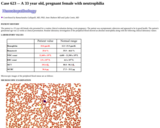
The patient is a 30-year old woman presenting with lower extremity edema during and following a pregnancy in 2005. She underwent a renal biopsy in 2005 which established the diagnosis of membranoproliferative glomerulonephritis-type III (Strife variant). She was treated with ACE inhibitors, but no steroids or cytoxic drugs over the next four years. In the initial biopsy, there were minimal chronicity changes with only one of twenty-six (1/26; 4%) glomeruli being globally sclerotic. Presently, the patient has nephrotic syndrome. Blood pressure is 110/70 mm Hg on anti-hypertensive medication. Pertinent laboratory data include: creatinine 0.6 mg/dl, BUN 13 mg/dl, urine protein 4.4 gm/24 hrs (4+ by dip stick), ANA-negative, ANCA-negative, hepatitis-B/C serology-negative, cryoglobulin screen-negative, urine sediment-inactive (no cells or casts). Kidneys are normal size by ultrasound. The clinical differential diagnosis includes: persistent/recurrent type-III membranoproliferative glomerulonephritis and a new glomerular cause of nephrotic syndrome.
(This case study was added to OER Commons as one of a …
(This case study was added to OER Commons as one of a batch of over 700. It has relevant information which may include medical imagery, lab results, and history where relevant. A link to the final diagnosis can be found at the end of the case study for review. The first paragraph of the case study -- typically, but not always the clinical presentation -- is provided below.)
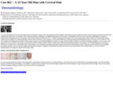
A 31 year old woman presented with worsening neck pain for 2 weeks. She was treated for muscle spasms at an urgent care clinic with muscle relaxants and narcotic pain medication, but had no relief of symptoms. On physical exam, she had focal tenderness in the midline at the base of the occiput and resistance to any motion of the neck. Routine laboratory examination was unremarkable. There was no associated history of trauma. CT scan showed a large, expansile, and destructive mass in the right clivus extending into the right petrous bone with associated right medulla effacement (Fig 1). Follow-up with MRI again showed a 5.0 x 3.0 x 3.0 cm expansile, destructive mass in the right clivus and along the right anterolateral margin of the foramen magnum/base of the skull (Fig 2).
(This case study was added to OER Commons as one of a …
(This case study was added to OER Commons as one of a batch of over 700. It has relevant information which may include medical imagery, lab results, and history where relevant. A link to the final diagnosis can be found at the end of the case study for review. The first paragraph of the case study -- typically, but not always the clinical presentation -- is provided below.)
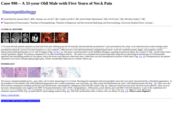
A 31-year-old man, with no family history of neuromuscular diseases, received muscle biopsy for slowly progressive muscle weakness. The first symptom he had was difficulty dorsiflexing his feet at age 20 years, which was followed by gradual development of gait disturbance. At age 27 years, he started using a handrail to climb up stairs. At age 28 years, he developed dysphagia for liquid, in addition to difficulty raising arms, which led him to have aspiration pneumonia later in the same year. At age approximately 30 years, he developed dyspnea on exertion. Arterial blood gas analysis revealed hypoxemia (65 mmHg, at room air) and hypercapnia (82 mmHg), with a vital capacity decreased to 780 ml, leading to the diagnosis of chronic type 2 respiratory failure, and non-invasive ventilation was started. At age 31 years, he was unable to walk without aid and required a wheelchair for long distances. Physical examination revealed moderate muscle weakness and atrophy in an asymmetric limb-girdle distribution, together with marked muscle atrophy in the tibialis anterior muscles and weakness in ankle dorsiflexion with Medical Research Council grade 1. Mild neck muscle weakness was also noted. Serum creatine kinase level was 375 IU/L (normal: <287 IU/L). Electromyography showed myopathic changes. Skeletal muscle CT demonstrated remarkable fat tissue replacement in the semitendinosus muscles (figure 1a, arrows) at the thigh level.
(This case study was added to OER Commons as one of a …
(This case study was added to OER Commons as one of a batch of over 700. It has relevant information which may include medical imagery, lab results, and history where relevant. A link to the final diagnosis can be found at the end of the case study for review. The first paragraph of the case study -- typically, but not always the clinical presentation -- is provided below.)
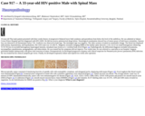
The patient is a 31 year old male with an unknown past medical history. The police initially found him naked, confused, and acting violently. He was taken to the emergency department, where his initial vitals and physical exam were significant for a temperature of 38.2oC, heart rate of 112 beats per minute, blood pressure of 156/108, diaphoresis, mydriasis, dried blood in his nares, skin pop marks on his arms, and no focal neurological deficits. Pertinent laboratory studies are included in Table 1.
(This case study was added to OER Commons as one of a …
(This case study was added to OER Commons as one of a batch of over 700. It has relevant information which may include medical imagery, lab results, and history where relevant. A link to the final diagnosis can be found at the end of the case study for review. The first paragraph of the case study -- typically, but not always the clinical presentation -- is provided below.)
A 31-year-old man was hospitalized due to retinitis and progressive personality changes that had started several weeks earlier. He was disorientated and had changes of affect with mood swings as well as signs and symptoms of dementia. Neuropsychologically the patient showed Balint's syndrome (paralysis of visual fixation, optic ataxia, and impairment of visual fixation) with anosognosia, visual and spatial agnosia, ideomotor and ideational apraxia, attention deficits and visual hallucinations. Electroencephalogram (EEG) showed non-specific abnormalities. The ophthalmological exam revealed retinitis with bilateral macular changes and partial atrophy of the optic nerves. Laboratory tests and cerebrospinal fluid (CSF) examinations were unremarkable. Magnetic Resonance Imaging (MRI) showed diffuse areas with high signal intensity in T2- weighted and FLAIR images involving periventricular and subcortical white matter of the occipital and parietal lobes. (Figures 1, 2). Furthermore, a focal 1cm mass lesion was detected in the sellar region. There was some clinical improvement with steroid treatment, but the patient refused further diagnostic procedures and was released to home.
(This case study was added to OER Commons as one of a …
(This case study was added to OER Commons as one of a batch of over 700. It has relevant information which may include medical imagery, lab results, and history where relevant. A link to the final diagnosis can be found at the end of the case study for review. The first paragraph of the case study -- typically, but not always the clinical presentation -- is provided below.)
A 32-year-old woman was referred to the Department of Neurosurgery, University of Zurich with a newly diagnosed supradiaphragmatic mass in the right paravertrebral region involving the paraspinal aspect of T10-12 and the T11 nerve root. The patient had noted a 2 year history of intermittent thoracic and upper abdominal pain radiating to the right side. No medical consultation was obtained during this period. After experiencing nausea and an episode of increased abdominal pain, which radiated to the right trunk and shoulder region, the patient presented to an emergency unit. There was no significant past medical history and the family history was negative regarding malignancies. On neurological examination no functional deficits were detected. MRI of the thoracic spine showed a circumscribed, right paravertebral and retrocrural multilobulated, encapsulated tumor measuring 46 x 42 x 58 mm. The tumor occupied the spinal canal between T10-T12, shifting the spinal cord to the left, protruding into the neuroforamina T10-T11 and infiltrated the 11th and 12th rib (Figs. 1a, 1b, 1c, 1d and 1e). Intra- and paraspinal resection of the tumor was performed in collaboration with a team from thoracic surgery. The infiltrated T11 nerve root was resected as well as the facet joints of T10/11 and T11/12 and the rib head of T11. A posterior decompression and instrumented fusion T10-T12 was performed. The tumor was highly pigmented with a dark brownish-black discoloration (Fig. 1f). The postoperative course was uneventful. Because of the diagnostic uncertainty, FDG-PET of the entire body, cranial MRI, fundoscopy, and gynecological examination were obtained, but no primary lesion was found. At 6 weeks clinical follow-up, the patient noted a significant decrease in thoracic and abdominal pain. Postoperative treatment consisted of adjuvant radiotherapy (33 x 2 Gy = 66 Gy). The MRI follow-up at 3 months after the operation showed no residual or recurrent tumor tissue in the thoracic spine (Figs.1g, 1h, 1i and 1j). A 3 month follow up FDG-PET of the entire body was performed showing 3 lesions in the abdominal fat, highly suspicious of metastasis (Fig. 1k).
(This case study was added to OER Commons as one of a …
(This case study was added to OER Commons as one of a batch of over 700. It has relevant information which may include medical imagery, lab results, and history where relevant. A link to the final diagnosis can be found at the end of the case study for review. The first paragraph of the case study -- typically, but not always the clinical presentation -- is provided below.)
This 32-year-old man without specific underlying disease suffered from intermittent headache for more than half a month. The pain was localized over left side temporal area and then transferred to left occipital area. It could be relieved with acetaminophen. However a severe headache episode, with repetitive vomiting, aroused him from sleep early one morning. He was sent to the emergency department with stable vital signs and clear consciousness. On examination, there was no anisocoria, limbs weakness, dysphasia, dysarthria, or palsy of cranial nerves.
(This case study was added to OER Commons as one of a …
(This case study was added to OER Commons as one of a batch of over 700. It has relevant information which may include medical imagery, lab results, and history where relevant. A link to the final diagnosis can be found at the end of the case study for review. The first paragraph of the case study -- typically, but not always the clinical presentation -- is provided below.)
A 32-year-old man presented with a 7-month history of headache and 2-month visual loss. Neurologic examination was unremarkable except for low visual acuity, worse in the left eye. Magnetic resonance imaging showed symmetrical cortical lesions in the midline, affecting predominantly both cingulate gyri and the upper corpus callosum. Lesions appeared multifocal, often limited to the cerebral cortex, confluent with speckled appearance, high signal intensity in T1-weighted images (Fig. 1), isointense in T2, strong contrast enhancement (Fig. 2), hypoperfusion with low regional cerebral blood flow values. Multivoxel spectroscopy showed increased choline/creatine ratio (2.97). There was prominent symmetrical edema of centrum semiovale. No changes were found in the leptomeninges.
(This case study was added to OER Commons as one of a …
(This case study was added to OER Commons as one of a batch of over 700. It has relevant information which may include medical imagery, lab results, and history where relevant. A link to the final diagnosis can be found at the end of the case study for review. The first paragraph of the case study -- typically, but not always the clinical presentation -- is provided below.)
NEUROPATHOLOGIC FINDINGS Brain sectioning revealed, apart from macroscopic findings easily identifiable as consequences of the subarachnoid hemorrhage in the past (multiple cortical infarcts, "coiled" basilar aneurysm etc.), three unusual yellowish microlesions in the left and right nucleus ruber (2mm and 4mm in diameter, respectively) and pons (<0.5mm in diameter). Histopathological examination showed that these lesions were of moderate cell density and had clearly defined, but irregularly borders (Figure 1). The Gomori silver stain revealed a well-established network of reticulin fibers surrounding these cells (Figure 2). The lesions consisted of ovoid to elongated cells that were positive for Vimentin, S100-protein (Figure 3) and myelin basic protein (MBP) (Figure 4) but did not react for 2',3'-Cyclic-Nucleotide 3'-Phosphodiesterase (CNP). These cells were intermingled with some lipid-laden macrophages. Within the lesions pre-existing axons seemed to be largely preserved as visualized with an antibody detecting neurofilament protein (NF) (Figure 5). The adjacent brain parenchyma showed reactive astrogliosis. The lesions did not react with antibodies staining for actin, desmin, EMA, synaptophysin, NeuN, Melan A, CD31, p53 or IDH-1. The proliferation index estimated with Ki-67 (MIB-1) was low (approximately 1%). It is worth mentioning that all three lesions were located in the proximity of small stage III infarctions.
(This case study was added to OER Commons as one of a …
(This case study was added to OER Commons as one of a batch of over 700. It has relevant information which may include medical imagery, lab results, and history where relevant. A link to the final diagnosis can be found at the end of the case study for review. The first paragraph of the case study -- typically, but not always the clinical presentation -- is provided below.)
A 32-year-old man was admitted to the neurosurgery department due to progressive dizziness and weakness of the feet for a month. An MRI revealed a well-demarcated mass measuring 60x35x25 mm, obstructing the fourth ventricle with dilatation of the lateral ventricles, third ventricle and the aqueduct. The lesion showed contrast enhancement with heterogeneous low signal intensity on T1WI (Figure 1) and mixed heterogeneous low and iso-signal intensity on T2WI (Figure 2). Several foci of cystic change were noted. A gross total resection was performed.
(This case study was added to OER Commons as one of a …
(This case study was added to OER Commons as one of a batch of over 700. It has relevant information which may include medical imagery, lab results, and history where relevant. A link to the final diagnosis can be found at the end of the case study for review. The first paragraph of the case study -- typically, but not always the clinical presentation -- is provided below.)
The patient is a 32 year old male who presented to the emergency department complaining of progressive fatigue and shortness of breath on exertion over a two to three month period. He denied any infection, bleeding or bruising. He reported consumption of one case of beer per day for ten years, but in the last four years had reduced his intake to one six-pack per day. Past medical history is significant for multiple gunshot wounds to the abdomen in the late 90s requiring resection of small bowel and colon, including the terminal ileum and cecum. This was followed by repair of a large ventral hernia in 2001. No outpatient medications and no known drug allergies. Physical exam showed mild scleral icterus, lungs clear to auscultation, heart regular rate and rhythm without murmurs, rubs, or gallops, abdomen with midline scar, liver palpable 2 cm below the left costal margin, and spleen not enlarged.
(This case study was added to OER Commons as one of a …
(This case study was added to OER Commons as one of a batch of over 700. It has relevant information which may include medical imagery, lab results, and history where relevant. A link to the final diagnosis can be found at the end of the case study for review. The first paragraph of the case study -- typically, but not always the clinical presentation -- is provided below.)
A 37-year-old woman presented with the eye sight declined repeatedly for eight months and became more and more serious. On clinical examination, she had developed blurred vision but no focal neurological signs. Laboratory analysis showed the following: red blood cell (RBC) count, 2.59×10¹²/L; hemoglobin, 88.0 g/L; mean corpuscular volume (MCV), 106.3 fl; mean corpuscular hemoglobin (MCH), 35.0 pg; mean corpuscular hemoglobin concentration (MCHC), 330.0g/L; red blood cell distribution width (RDW), 29.0%; white blood cell (WBC) count, 15.9×10⁹/L; 55% orthochromatic erythroblasts; platelet count, 92.0×10⁹/L.
(This case study was added to OER Commons as one of a …
(This case study was added to OER Commons as one of a batch of over 700. It has relevant information which may include medical imagery, lab results, and history where relevant. A link to the final diagnosis can be found at the end of the case study for review. The first paragraph of the case study -- typically, but not always the clinical presentation -- is provided below.)
A 33 year-old patient with no relevant clinical history presented in August 2007 with right paravertebral rigidity. Patient had previously undergone kinetic physical therapy and been prescribed anti-inflammatory medication. In December 2007, patient presented to Neurology with right brachial paresis (3/5), positive osteotendinous reflexes, and cervical pain. MRI (Figure 1) showed a hyperintense intraspinal lesion on T2 sequence with contrast enhancement between C2-C7. Diffuse thickening of the spinal cord was also appreciated. January 2008: CSF showed high protein content, but was negative for oligoclonal bands. PCR for HSV and TB were also negative, as was serum NMO-IgG. Evoked potentials showed bilateral increase in latency (117.5 OD-115.5 OI). The condition was interpreted as myelitis. Patient began treatment with methylprednisolone (5 pulses), in spite of which medical condition worsened, developing nystagmus, muscle spasms, and progression of motor deficit with altered osteotendinous reflexes (February-March 2008). In March-April 2008, bilateral hand paresthesias, incontinence, severe right hemiparesis and sensory alterations were observed. In April 2008, patient was brought to our institution presenting bradycardia, hypotension (shock), and respiratory failure. Physical exam revealed flaccid tetraplegia, generalized areflexia, reactive isochoric pupils and horizontal nystagmus. Imaging revealed spinal lesion had increased in size and extended to the medulla (Figure 2). Anoxic-ischemic encephalopathy was diagnosed. Patient died in May 2008.
(This case study was added to OER Commons as one of a …
(This case study was added to OER Commons as one of a batch of over 700. It has relevant information which may include medical imagery, lab results, and history where relevant. A link to the final diagnosis can be found at the end of the case study for review. The first paragraph of the case study -- typically, but not always the clinical presentation -- is provided below.)
A 33-year-old male patient reported on head and neck pain radiating into the left maxilla, that had already persisted for 5 years presented in the clinic, as he experienced an acute neuralgic pain provoked by physical activity (VAS 8/10 points) as a new symptom. MRI showed a left-sided hypointense, polyglobulated tumor in the left cerebello-pontine angle, with irregular contrast enhancement and hypointensity on T1 and T2 images (Figs. 1a, 1b, 1c). The tumor exerted pressure on the medulla oblongata, resulting in perifocal edema. By cranial CT (Fig. 1d) the tumor had a strong hyperdense signal. The patient complained of no other neurological deficits. The tumor was extirpated microneurosurgically using electro-physiological monitoring via retromastoidal approach and semisitting position of the patient. Since the lesion was adjacent to the brain stem, a thin layer was left (intraoperative pictures of the tumor: Figs. 1e, 1f). Postoperatively, the patient sustained a new facial and glossopharyngeal palsy, which considerably improved at 3 months' follow-up.
(This case study was added to OER Commons as one of a …
(This case study was added to OER Commons as one of a batch of over 700. It has relevant information which may include medical imagery, lab results, and history where relevant. A link to the final diagnosis can be found at the end of the case study for review. The first paragraph of the case study -- typically, but not always the clinical presentation -- is provided below.)
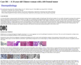
A 33-year-old Chinese woman presented with intermittent slurring of speech, dysphasia together with right upper limb and facial weakness for two months with gradual worsening of the symptoms. Physical examination found decreased pin-prick sensation over right C6 to C8 dermatome and impaired proprioception in right hand. CT scan with contrast showed a well-demarcated contrast-enhancing left frontal tumor measuring 4.5 x 4.2 x 3 cm with perilesional edema and slight mass effect. Cystic changes were observed. The tumor was close to the cortical surface but not connected to the meninges (Figure 1). Surgical exploration found a non-encapsulated, well-circumscribed, vascularized tumor in the left frontal lobe. Tumor debulking under intraoperative cerebral function monitoring was performed. Around 95% of tumor was removed but complete excision could not be achieved due to significant decrease in amplitude of the brain motor evoked potentials. The patient recovered well after the operation with complete restoration of the function in the precentral and postcentral gyri as well as Broca's area.
(This case study was added to OER Commons as one of a …
(This case study was added to OER Commons as one of a batch of over 700. It has relevant information which may include medical imagery, lab results, and history where relevant. A link to the final diagnosis can be found at the end of the case study for review. The first paragraph of the case study -- typically, but not always the clinical presentation -- is provided below.)
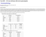
33-year-old Thai male patient presented with three weeks history of progressive bilateral lower limb weakness and paresthesia from below the level of the umbilicus. He was admitted at Sakaeo Crown Prince Hospital and first diagnosed with HIV/AIDS. He did not receive antiretroviral drugs before. Neurological examination showed loss of motor power of both lower extremities. Sensory loss was detected in all dermatomes below T10. Areflexia was observed in both legs. The meningeal sign was negative. The other systems revealed no remarkable change. The blood test displayed mild anemia, hyponatremia, and hypokalemia. His CD4 count was 14 cells/?L. Magnetic resonance imaging (MRI) of the lumbar spine showed a 1.4x1.3x1.0 cm round homogenous enhancing, T1/T2 Wi-low-to-iso/mild heterogenous hypersignal intensity, intradural mass lesion at L1 vertebral body level [Figure 1a]. Radiologically, the differential diagnosis included schwannoma, myxopapillary ependymoma, paraganglioma, and tuberculoma. He was treated but his symptoms did not improve. Few weeks later, he was referred to a specialist at our hospital. Surgeon performed lumbar laminectomy with gross total resection of tumor. Postoperatively, he developed progressive dyspnea with clinical suspicion for Pneumocystis jiroveci pneumonia (PJP) infection. He was treated but his clinical symptom got worse. Finally, he developed respiratory failure and expired two weeks after operation.
(This case study was added to OER Commons as one of a …
(This case study was added to OER Commons as one of a batch of over 700. It has relevant information which may include medical imagery, lab results, and history where relevant. A link to the final diagnosis can be found at the end of the case study for review. The first paragraph of the case study -- typically, but not always the clinical presentation -- is provided below.)
A 33-year-old male presented with a 2-week old uncomfortable mass within his left testicle. His serum -hCG was elevated (345.2 mIU/mL). Ultrasound examination demonstrated a heterogeneous, hypoechoic mass. The patient subsequently underwent left radical orchiectomy and epididymectomy. Two weeks after the operation -hCG level was less than 2 mIU/ml. Clinical and radiological staging was negative.
(This case study was added to OER Commons as one of a …
(This case study was added to OER Commons as one of a batch of over 700. It has relevant information which may include medical imagery, lab results, and history where relevant. A link to the final diagnosis can be found at the end of the case study for review. The first paragraph of the case study -- typically, but not always the clinical presentation -- is provided below.)
The patient is a 33 year old female who presented for a routine clinical evaluation during a twin pregnancy. The patient was asymptomatic otherwise and appeared to be in good health. The patient's gestational age was 22 weeks at clinical presentation. Routine laboratory investigation of the peripheral blood showed an absolute neutrophilia along with the following clinical laboratory values:
(This case study was added to OER Commons as one of a …
(This case study was added to OER Commons as one of a batch of over 700. It has relevant information which may include medical imagery, lab results, and history where relevant. A link to the final diagnosis can be found at the end of the case study for review. The first paragraph of the case study -- typically, but not always the clinical presentation -- is provided below.)
A 31-year-old, previously healthy woman experienced a new-onset generalized seizure with subsequent right-sided weakness in the 37th week of her pregnancy. History revealed no significant nausea or vomiting (other than that associated with the first trimester of pregnancy) and no prior history of seizures. The patient did, however, report a history of slowly-increasing weakness in her right leg over the past several months. Routine prenatal care had been uneventful and negative for gestational diabetes or hypertension.
(This case study was added to OER Commons as one of a …
(This case study was added to OER Commons as one of a batch of over 700. It has relevant information which may include medical imagery, lab results, and history where relevant. A link to the final diagnosis can be found at the end of the case study for review. The first paragraph of the case study -- typically, but not always the clinical presentation -- is provided below.)
A 33 year old female patient presented in June 2009 with progressive loss of vision for the last 12 months plus occasional episodes of headache. In the last 3 months her vision deteriorated even further with progressive temporal amaurosis, and she started having depression with episodes of extreme anxiety. She developed amenorrhea since her last pregnancy 8 years ago with episodes of galactorrhea. CT scan (Figure 1) showed a mass lesion in the sellar region suggestive of meningeoma or pituitary adenoma. She received bromocriptine 2.5mg/day with no reduction of the lesion. She developed panhypopituitarism with normal levels of prolactin. Further MRI showed a large lesion infiltrating cavernous sinuses and compressing the optic nerve. Transsphenoidal surgical treatment was performed. Two months after the first operation she returned to hospital with complete bilateral amaurosis and a new MRI showed residual lesion. A new sub-frontal neurosurgery was performed and most of the lesion was resected. The patient developed diabetes insipidus but remains under control.
No restrictions on your remixing, redistributing, or making derivative works. Give credit to the author, as required.
Your remixing, redistributing, or making derivatives works comes with some restrictions, including how it is shared.
Your redistributing comes with some restrictions. Do not remix or make derivative works.
Most restrictive license type. Prohibits most uses, sharing, and any changes.
Copyrighted materials, available under Fair Use and the TEACH Act for US-based educators, or other custom arrangements. Go to the resource provider to see their individual restrictions.