(This case study was added to OER Commons as one of a …
(This case study was added to OER Commons as one of a batch of over 700. It has relevant information which may include medical imagery, lab results, and history where relevant. A link to the final diagnosis can be found at the end of the case study for review. The first paragraph of the case study -- typically, but not always the clinical presentation -- is provided below.)
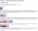
A 34 year-old otherwise healthy man presented with new onset headaches, mild confusion, and subtle left-sided weakness. An MRI scan showed a large heterogeneous 8.9cm x 6.0cm right fronto-temporo-parietal mass with nodular enhancement, significant surrounding edema, and 15mm of midline shift. He underwent an urgent right craniotomy, and began appropriate therapy. Unfortunately, within one week of completion of this therapeutic regimen, his symptoms worsened. He was found to have multiple new bilateral dural-based nodular enhancing lesions on repeat brain MRI. No spinal abnormalities were found. He underwent resection of one of the new lesions. The patient was initiated on a new treatment plan, but unfortunately, neuroimaging revealed further progression of disease (Figure 1). He passed away 5 months and 1 day after diagnosis.
(This case study was added to OER Commons as one of a …
(This case study was added to OER Commons as one of a batch of over 700. It has relevant information which may include medical imagery, lab results, and history where relevant. A link to the final diagnosis can be found at the end of the case study for review. The first paragraph of the case study -- typically, but not always the clinical presentation -- is provided below.)
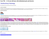
The patient is a 34 year-old morbidly obese man who presented with abdominal pain and dysuria. A CT scan was performed and revealed a small nodule on the dome of his bladder. Cystoscopy was performed and demonstrated a 2-3 cm tumor at the bladder dome with purulent discharge. A partial cystectomy was performed.
(This case study was added to OER Commons as one of a …
(This case study was added to OER Commons as one of a batch of over 700. It has relevant information which may include medical imagery, lab results, and history where relevant. A link to the final diagnosis can be found at the end of the case study for review. The first paragraph of the case study -- typically, but not always the clinical presentation -- is provided below.)
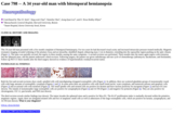
This 34 year-old man presented with a five month complaint of bitemporal hemianopsia. For two years he had decreased visual acuity and increased intraocular pressure treated medically. Magnetic resonance imaging revealed widening of the pituitary fossa and an intrasellar, dumbbell shaped, enhancing mass 3 cm in diameter, extending into the suprasellar region pushing on the optic chiasm (Figure 1). The mass was partially excised, but regrew in three months causing the same symptoms. A second excision was performed. Three months later the tumor again regrew with extension into the sphenoid sinus, and the patient underwent a third excision. Following surgery, the patient was given radiotherapy and one cycle of chemotherapy (adriamycin, dacabarzine, and ifosfamide). Follow-up PET-CT three months after the third surgery showed no evidence of hypermetabolic residual/recurrent tumor.
(This case study was added to OER Commons as one of a …
(This case study was added to OER Commons as one of a batch of over 700. It has relevant information which may include medical imagery, lab results, and history where relevant. A link to the final diagnosis can be found at the end of the case study for review. The first paragraph of the case study -- typically, but not always the clinical presentation -- is provided below.)
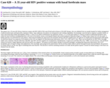
The patient was a 35-year-old African American woman with HIV/AIDS (CD4 count 95/ml) and no history of HAART therapy, who was admitted from an outside hospital for further management of altered mental status and hypercarbic respiratory failure. Upon admission, the patient was unresponsive and ventilator dependent. On neurologic exam she had no response to painful stimuli, as well as decreased deep tendon and pupillary reflexes. A chest X-ray failed to demonstrate any pulmonary lesions. A CT of the head without IV contrast revealed an infiltrating hypodense mass within the basal forebrain extending inferiorly to the level of the basilar cisterns, involving the thalami and cerebral peduncles bilaterally (Figure 1). Diffuse brain volume loss was also noted. A lumbar puncture was performed for CSF analysis, which showed lymphocytic pleocytosis (56 nucleated cells/ml with 92% lymphocytes), low glucose (23 mg/dl) and elevated protein (208 mg/dl). An infectious disease was suspected. PCR analysis of the CSF was negative for Mycobacterium tuberculosis, HSV, VZV, CMV and JC virus. There was low positivity for EBV DNA. Serologic studies were consistent with past infections with EBV and CMV and were negative for toxoplasmosis, histoplasmosis, Cryptococcus and West Nile virus. VDRL was non-reactive. CSF and blood cultures for bacteria, fungal organisms and acid fast bacilli were negative. The patient received empiric therapy for bacterial meningitis, including Mycobacterium tuberculosis, HSV/VZV encephalitis and coverage for fungi with amphotericin. Her neurologic status progressively deteriorated. She lost all deep tendon reflexes and her left pupil became fixed and dilated. The presence of corneal reflexes was the only sign of remaining brain stem function. The decision to end life sustaining treatment was made and the patient expired. Consent for limited autopsy of the brain was obtained from the family.
(This case study was added to OER Commons as one of a …
(This case study was added to OER Commons as one of a batch of over 700. It has relevant information which may include medical imagery, lab results, and history where relevant. A link to the final diagnosis can be found at the end of the case study for review. The first paragraph of the case study -- typically, but not always the clinical presentation -- is provided below.)
A previously healthy, 35-year-old man presented with a four-week history of painful swelling on his right forehead. CT imaging showed a solitary, 1 cm osteolytic lesion within the diploic space of the right frontal bone, involving the inner and outer tables (Figure 1). On MRI, the lesion was heterogeneously enhancing and extended into subgaleal tissue and epidural space. It was resected and the patient was discharged in good condition.
(This case study was added to OER Commons as one of a …
(This case study was added to OER Commons as one of a batch of over 700. It has relevant information which may include medical imagery, lab results, and history where relevant. A link to the final diagnosis can be found at the end of the case study for review. The first paragraph of the case study -- typically, but not always the clinical presentation -- is provided below.)
The patient is 35-year-old previously healthy native African male, who moved to the United States two years ago. He presented to an outside facility with a chief complaint of anal and rectal bleeding. Laboratory values are unremarkable, and include: hemoglobin 13.1 gm/dL, hematocrit 39%, white blood cell count 6,000 cell/cc, eosinophils 2.9% (normal 0.2-6.0%), platelets 224, calcium 9.2 mg/dL, albumin 3.7 g/dL, glucose 94 mg/dL, AST 51 IU/L, ALT 45 IU/L, alkaline phosphatase 47 IU/L, total bilirubin 0.5 mg/dL, sodium 135 mmol/L, potassium 4 mmol/L, chloride 110 mmol/L, blood urea nitrogen 9 mg/dL, and creatinine 1.1 mg/dL. Colonoscopy revealed thrombosed external hemorrhoids and mild erythema in the rectal area. There were also small, non-bleeding internal hemorrhoids. The terminal ileum appeared normal. Clinically, the differential diagnosis included colonoscopy preparation-related changes and a mild colitis. The colonoscopy findings were sufficient to explain mild hematochezia, and fiber supplementation with repeat screening colonoscopy in ten years was recommended.
(This case study was added to OER Commons as one of a …
(This case study was added to OER Commons as one of a batch of over 700. It has relevant information which may include medical imagery, lab results, and history where relevant. A link to the final diagnosis can be found at the end of the case study for review. The first paragraph of the case study -- typically, but not always the clinical presentation -- is provided below.)
The patient is a previously healthy 35-year-old man who was admitted to our hospital for evaluation after experiencing vomitus, dizziness and headache for one month. A neurological examination elicited no abnormalities. CT and MR imaging were performed and revealed a 3.5 x 3.2 cm solitary, well-delineated, extra-axial midline mass arising from the frontal falx cerebri. The lesion was isointense to the cortex on T2 (Figure 1) and hypointense on T1, with a marked homogenous enhancement after intravenous contrast administration (Figures 2 and 3). Based on these findings, the radiological diagnosis was meningioma of the falx. At surgery, the tumour appeared as an extra-axial lesion and was removed via a left midline frontal craniotomy. The excision was macroscopically complete.
(This case study was added to OER Commons as one of a …
(This case study was added to OER Commons as one of a batch of over 700. It has relevant information which may include medical imagery, lab results, and history where relevant. A link to the final diagnosis can be found at the end of the case study for review. The first paragraph of the case study -- typically, but not always the clinical presentation -- is provided below.)
A 35-year-old woman presented for neurological evaluation after a single episode of speech disruption consisting of word finding difficulty and a partial motor seizure followed by one hour of confusion. A head MRI scan was performed, which showed a 2.7 by 2.0 by 2.5 cm dural-based contrast enhancing lesion along the left posterior temporal lobe (Figure 1). An EEG at the time of presentation was negative, but the patient was placed on prophylactic Dilantin therapy. She was referred for neurosurgical evaluation, and a recommendation was made for surgical resection. A left parietal-temporal craniotomy was performed to resect the tumor. At surgery, a firm dural-based lesion was encountered with no evidence of brain invasion or involvement, and a gross total resection was performed. The patient tolerated the procedure well, and was discharged home on postoperative day three. The patient had no further seizures or episodes of speech difficulty, and was tapered off of her anticonvulsants six months following surgery, with a second EEG negative at that time. Follow-up MRI scans at 6 and 12 months showed no evidence of tumor recurrence, and she continues to have a normal neurological exam.
(This case study was added to OER Commons as one of a …
(This case study was added to OER Commons as one of a batch of over 700. It has relevant information which may include medical imagery, lab results, and history where relevant. A link to the final diagnosis can be found at the end of the case study for review. The first paragraph of the case study -- typically, but not always the clinical presentation -- is provided below.)
A 35 year old woman was admitted to our hospital with symptoms and signs of acromegaly that started one year earlier. Physical examination detected clubbing of her fingers, enlargement of her jaws, a carpal tunnel syndrome along with joint and neck pain. Laboratory findings showed hypothyroidism, secondary hypogonadism, diabetes mellitus and anemia. Cranial MRI revealed an intra- and suprasellar mass lesion with radiological signs of a chiasma syndrome. She was treated with a somatostatin therapy. Although the clinical symptoms improved over the next three months, Insulin like Growth Factor-1 levels ( a marker for acromegaly) continued to be at pathological levels. The patient therefore underwent a transphenoidal resection of the intrasellar mass.
(This case study was added to OER Commons as one of a …
(This case study was added to OER Commons as one of a batch of over 700. It has relevant information which may include medical imagery, lab results, and history where relevant. A link to the final diagnosis can be found at the end of the case study for review. The first paragraph of the case study -- typically, but not always the clinical presentation -- is provided below.)
A 35-year-old G2P2, presented at nine-months post-partum to her gynecologist for increased left labial swelling over a 2-month period. Pertinent medical history included two spontaneous vaginal deliveries, menorrhagia with known uterine fibroids, and no abnormal Pap smears. Physical examination at that time demonstrated a painless 7 cm mass in the area of the left Bartholin gland. An incision and drainage performed by the primary gynecologist revealed expression of gelatinous material. The mass recurred over the next two months, and the patient transferred her care to our institution. A radical vulvectomy with additional deep margins was performed at our institution.
(This case study was added to OER Commons as one of a …
(This case study was added to OER Commons as one of a batch of over 700. It has relevant information which may include medical imagery, lab results, and history where relevant. A link to the final diagnosis can be found at the end of the case study for review. The first paragraph of the case study -- typically, but not always the clinical presentation -- is provided below.)
A 35-year-old woman presented with one month's history of progressive bilateral leg weakness and altered sensation. There had been no pain. She had noted urinary frequency and constipation in the previous two weeks. On examination, the patient had diffuse lower extremity weakness (2-3/5), with a T6 sensory level to pain and temperature sensation. Proprioception was preserved. Post-void residuals exhibited urinary retention. There was sacral hypesthesia and decreased rectal tone. She was mildly hyperreflexic (3/4) at the knees and ankles without clonus; both great toes were upgoing. A T1-weighted MRI demonstrated a T4-5 intradural mass ventral to the spinal cord (Fig. 1A), with an enhancing dural tail (Fig. 1B), consistent with meningioma. The lesion was dark on T2-weighted images (Fig. 1C).
(This case study was added to OER Commons as one of a …
(This case study was added to OER Commons as one of a batch of over 700. It has relevant information which may include medical imagery, lab results, and history where relevant. A link to the final diagnosis can be found at the end of the case study for review. The first paragraph of the case study -- typically, but not always the clinical presentation -- is provided below.)
We present a 36-year-old cognitively intact male, with history of work-related solvent exposure who consulted for headache, vomiting and fever in September 2009. Symptoms persisted in spite of antibiotic therapy. CT scan showed a hypodense image in the left parietal lobe. MRI revealed a parietal lesion hypointense on T1, hyperintense on T2, and FLAIR, without mass effect (Fig. 1a). Thoracic CT scan showed bi-apical interstitial infiltrates. Standard TB treatment was indicated along with steroids, obtaining a partial clinical response. Two months later, following steroid tapering, significant clinical deterioration was observed with fever, headaches and nausea. Patient was re-evaluated showing no changes on MRI. PCR testing for several microorganisms was negative. Differential diagnoses considered were: recurrent Meningitis NOS vs. vasculitis vs. ADEM (Acute Disseminated Encephalomyelitis). A new course of methylprednisolone was administered; after which patient requested voluntary discharge. In December 2009, patient returned with persistent fever and headache. A methylprednisolone bolus was administered and partial, transient response observed. At this time, the diagnosis considered was ADEM refractory to steroids, and treatment was switched to immunoglobulin therapy, which led to a complete clinical response. Once again, patient requested voluntary discharge, but had to be re-admitted later, due to headache, fever, mental confusion and aphasia in January 2010. New CSF PCRs ruled out viral diseases and TB. Other differential diagnoses considered included: Marburg's disease, CNS lymphoma and Neuro-Behçet's disease. The patient then suffered rapid clinical decline, developing limb weakness, somnolence and hyponatremia and died in January 2010.
(This case study was added to OER Commons as one of a …
(This case study was added to OER Commons as one of a batch of over 700. It has relevant information which may include medical imagery, lab results, and history where relevant. A link to the final diagnosis can be found at the end of the case study for review. The first paragraph of the case study -- typically, but not always the clinical presentation -- is provided below.)
A 36-year-old woman was referred to our unit for subacute onset (within 2 months) of right eye ptosis and unreactive mydriasis. The patient complained of photophobia, right ocular dryness and progressive inability in focusing on images. A neurological examination at a local emergency room found incomplete III cranial nerve palsy. A CT scan was normal. The right fundus oculi evaluation with optic coherence tomography (OCT) showed papillary edema. Visual fields examination showed blind spot enlargement in the right eye. An MRI showed slightly T2 hyperintense tissue enlarging Meckel's cave and involving the carotid artery (Figs. 1a, 1b). The mass involved the right temporal dura, the cavernous sinus and extended into the right orbit through the superior orbital fissure (Figs. 1c, 1d). The right superior orbital fissure enlargement was well documented by an orbital CT scan (3D reconstruction, Fig. 1e). After Gd administration, it presented intense and homogenous enhancement (Figs. 1f, 1g) leading to the suspect of a lymphomatous or granulomatous lesion (neurosarcoidosis, Tolosa-Hunt syndrome). During the admission the patient underwent a complete serological investigation for infectious diseases and autoimmune disorders which resulted negative except for a slightly increased ACE level (57, n.v. = 8-52). Blood Calcium and parathormone (PTH) level were normal. The cerebrospinal fluid (CSF) biochemical analysis did not show any significant alteration and atypical lymphocytes were not found. Visual evocated potential showed normal latencies with increased cortical responses amplitudes. A chest CT scan showed only a small ground glass area in the apical segment of the right inferior pulmonary lobe. There were no radiological signs of interstitial abnormalities or mediastinal lymphadenopathy. A high dose corticosteroids (CCS) cycle was tempted without significant clinical improvement. After few days, the right eye ptosis consistently worsened and the patient developed right lateral gaze diplopia and hypoesthesia in the second trigeminal branch (V2) territory. After the CCS cycle a second MRI was repeated showing a diffuse increase of the lesion enhancement after Gd administration. It also involved the right muscle cone and the sheets of the right optic and trigeminal nerve through the oval foramen thus compressing the right carotid syphon. The mass infiltrated the left internal pterygoid muscle and extended in the left part of upper nasopharynx. Therefore, the patient underwent endoscopic endonasal biopsy performed through a bilateral anterior sphenoidectomy and a right transmaxillo-pterygoid approach to the pterygopalatine fossa.
(This case study was added to OER Commons as one of a …
(This case study was added to OER Commons as one of a batch of over 700. It has relevant information which may include medical imagery, lab results, and history where relevant. A link to the final diagnosis can be found at the end of the case study for review. The first paragraph of the case study -- typically, but not always the clinical presentation -- is provided below.)
The patient is an approximately 30-year-old primigravida with an intrauterine dichorionic-diamniotic twin pregnancy. Prenatal ultrasound at 30 weeks gestation showed a multi-cystic mass without fetal parts suspicious for hydatidiform mole (twin A), and one live fetus with normal anatomy (twin B) with a corresponding enlarged, cystic placenta (placenta B). At 36 weeks gestation, the mother developed preeclampsia and preterm labor and delivered one live, morphologically normal female infant (twin B) weighing in the less than tenth percentile. The infant was transferred to the NICU after delivery for low birthweight and prematurity.
(This case study was added to OER Commons as one of a …
(This case study was added to OER Commons as one of a batch of over 700. It has relevant information which may include medical imagery, lab results, and history where relevant. A link to the final diagnosis can be found at the end of the case study for review. The first paragraph of the case study -- typically, but not always the clinical presentation -- is provided below.)
A 36-year-old man, with personal history of a right parietal cranial injury, was admitted for an evaluation of a right parietal mass that had slowly increased in the nine years prior to this presentation. General examination disclosed nothing. Skull examination revealed a palpable pulsatile and soft swelling. Routine laboratory tests were unremarkable. Computed tomography (CT) and magnetic resonance imaging (MRI) of the head were obtained to delineate the large mass in the parietal region. MRI showed a prominent extracranial scalp vein in direct communication with the superior sagittal sinus, through a right parietal bone defect. CT scan showed an old fracture with osteomeningeal breach. There was no parenchymal abnormality. The lesion was heterogeneously intense on T2- (Figure 1), T2-Flair (Figure 2) and T1-weighted images (Figure 3). Doppler sonography found vascular structures composing this lesion. Due to the worsening pain at the site of her swelling, a total resection was performed. Intra-operatively, the lesion was defined by the surgeon as a blood-filled sac within the pericranium and directly overlying the bone suggesting an angioma.
(This case study was added to OER Commons as one of a …
(This case study was added to OER Commons as one of a batch of over 700. It has relevant information which may include medical imagery, lab results, and history where relevant. A link to the final diagnosis can be found at the end of the case study for review. The first paragraph of the case study -- typically, but not always the clinical presentation -- is provided below.)
FD is a 36-year-old white woman with a history of an inherited genetic disease, diagnosed in 1999 when she was found to have a low leukocyte alpha-galactosidase enzyme level. She also has a remote history of headaches, tinnitus, vertigo, acroparesthesias, and angiokeratomas. There is extensive family involvement with this particular genetic disease, and those affected include her father, her teenage son, and two sisters. Her father died at age 49 from complications of cardiovascular and renal disease. The patient has no reported history of diabetes or hypertension; her recent blood pressure is 112/74 mmHg. An echocardiogram showed a normal left ventricular size and an ejection fraction of 60%. Pertinent laboratory values include: BUN 13 mg/dL, creatinine 0.6 mg/dL, hemoglobin 13.2 gm/dL, hematocrit 36.6%, and glucose 81 mg/dL. Urine analysis shows trace protein with no cells. The patient has no peripheral edema. She had microalbuminuria with urine albumin 4.2 mg/dL.
(This case study was added to OER Commons as one of a …
(This case study was added to OER Commons as one of a batch of over 700. It has relevant information which may include medical imagery, lab results, and history where relevant. A link to the final diagnosis can be found at the end of the case study for review. The first paragraph of the case study -- typically, but not always the clinical presentation -- is provided below.)
This patient is a 36 year-old male who is 216 days status post-kidney transplantation due to hypertension induced kidney failure. He had high creatinine (Cr) values with difficulties controlling his serum Cr which reached a value of 3.1 mg/dl. At that time, the patient was found to shed BK virus in his urine (BKV DNA: 7.7E05 copies/ml), however, no BKV DNA was detected in plasma. A renal allograft biopsy was performed. (Figure 1)
(This case study was added to OER Commons as one of a …
(This case study was added to OER Commons as one of a batch of over 700. It has relevant information which may include medical imagery, lab results, and history where relevant. A link to the final diagnosis can be found at the end of the case study for review. The first paragraph of the case study -- typically, but not always the clinical presentation -- is provided below.)
A 36-year old male presented to the hospital with an ear infection. Laboratory studies drawn in the emergency department demonstrated a pancytopenia including neutropenia. He was admitted to the hospital where he underwent an extensive workup including viral testing for hepatitis B, hepatitis C, and HIV, all of which were negative. Imaging of the sinuses was negative for acute or chronic sinusitis. His past medical history was significant for testicular cancer diagnosed at the age of 23. He is status post left orchiectomy and radiation therapy. He was in his usual state of health until approximately 3 months ago when he started having episodes of night sweats. Physical examination was significant for splenomegaly that measured approximately 3 fingerbreadths below the costal margin. The peripheral blood smear demonstrated anisocytosis, ovalocytes, polychromasia and tear drops. Atypical lymphoid cells were identified with rounded nuclear contours and hairy cytoplasmic projections (Figure 1). Platelets were reduced.
(This case study was added to OER Commons as one of a …
(This case study was added to OER Commons as one of a batch of over 700. It has relevant information which may include medical imagery, lab results, and history where relevant. A link to the final diagnosis can be found at the end of the case study for review. The first paragraph of the case study -- typically, but not always the clinical presentation -- is provided below.)
The patient is a 36 year-old African American male with a history of end stage renal disease secondary to IgA nephropathy. He underwent kidney transplantation in 2001, which subsequently failed in 2005 due to rejection. Allograft nephrectomy was performed in 2006 because of hematuria. He had been on peritoneal dialysis prior to his transplantation and wished to resume this method of renal replacement after the failure of his transplant. A dialysis catheter was placed in November 2005. Three months later, he presented with abdominal pain and cloudy peritoneal drainage. On physical exam, he was afebrile with normal vital signs. He had peri-umbilical tenderness with guarding and rebound tenderness. Peripheral white blood cell count was 10,600/cu mm with a normal differential. CT scan of the abdomen showed no free or loculated fluid collections. A sample of peritoneal fluid was sent for culture (Figs. 1, 2 and 3)
(This case study was added to OER Commons as one of a …
(This case study was added to OER Commons as one of a batch of over 700. It has relevant information which may include medical imagery, lab results, and history where relevant. A link to the final diagnosis can be found at the end of the case study for review. The first paragraph of the case study -- typically, but not always the clinical presentation -- is provided below.)
A 37 year old female from Brazil presented with paresthesia and cramping in the left calf. She also reported transient loss of strength in the left arm. Neurological examination revealed paresthesia on the entire left side of her body. No clinical abnormality was noted on physical examination, including breast and lymph node examination. During hospitalization, she presented sensorimotor seizure of the left arm and leg. Cerebral MRI showed a rolandic right lesion of postcentral gyrus with FLAIR hypersignal (Figure 1), heterogeneous enhancement and edema around the lesion (Figures 1 and 2). Laboratory tests, including blood cell count and CSF puncture, did not find any abnormality. Cerebral biopsy for diagnosis was performed.
No restrictions on your remixing, redistributing, or making derivative works. Give credit to the author, as required.
Your remixing, redistributing, or making derivatives works comes with some restrictions, including how it is shared.
Your redistributing comes with some restrictions. Do not remix or make derivative works.
Most restrictive license type. Prohibits most uses, sharing, and any changes.
Copyrighted materials, available under Fair Use and the TEACH Act for US-based educators, or other custom arrangements. Go to the resource provider to see their individual restrictions.