(This case study was added to OER Commons as one of a …
(This case study was added to OER Commons as one of a batch of over 700. It has relevant information which may include medical imagery, lab results, and history where relevant. A link to the final diagnosis can be found at the end of the case study for review. The first paragraph of the case study -- typically, but not always the clinical presentation -- is provided below.)
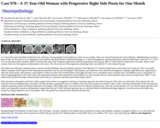
A 37-year-old woman had bilateral blurred vision with hazy vision gradually from periphery to center for two months. There was mixed horizontal and vertical diplopia. Ophthalmologic treatment did not help. We she came to us, an Humphrey visual field test disclosed bilateral temporal hemianopia. CT showed hyperdensity and heterogeneously enhanced sellar lesion with size 2.57 x 1.96 x 3.63 cm and supra-sellar extension. MRI T1 showed sella tumor with isointense signal mixed with the hyperintense part (Fig 1a). MRI T2 scans showed compression of the optic chiasm. With contrast, there was heterogeneous enhancement in the tumor. The clivus showed a heterogenously enhancing lesion was noted on T1 with contrast (Fig 1b).
(This case study was added to OER Commons as one of a …
(This case study was added to OER Commons as one of a batch of over 700. It has relevant information which may include medical imagery, lab results, and history where relevant. A link to the final diagnosis can be found at the end of the case study for review. The first paragraph of the case study -- typically, but not always the clinical presentation -- is provided below.)
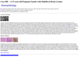
A 36-year old right-handed female patient in the 21st week of pregnancy presented to the Neurology emergency department due to double vision, hypoesthesia on the right side of her face, headache, nausea and weight loss. The symptoms had started 4 months previously, when she was going through an IVF (in vitro fertilisation) procedure, with nausea and weight loss. She became pregnant a few weeks after the symptoms started. Neurological examination revealed right abducens nerve palsy and sensory loss in the area of the right maxillary and mandibular nerve. MRI, T1 weighted sequence, showed multiple lesions in the ventricular system, and one in the region of the right trigeminal nerve, which could have been metastases (Figures 1A and 1B - arrows pointed the tumors). The complete blood count showed mild anaemia. A lumbar puncture was performed, which revealed normal opening pressure, mild pleocytosis (6 leucocytes) and slightly elevated proteins. There were no malignant cells in the CSF, chest X-ray, and abdomen and breast ultrasound were unremarkable. The neurosurgeon suggested a biopsy of the lesions next to the trigeminal nerve but only after the delivery. The patient did not consider abortion. Her neurological condition slowly deteriorated and a caesarean section was performed in the 30th week of pregnancy, followed immediately by a biopsy of the lesion in the region of the right trigeminal nerve. She gave birth to a girl weighed 1400g. After the delivery, the patient's condition started to deteriorate at a more rapid pace, with a worsening of headache with vomiting, disorientation and gait instability. A head CT scan showed obstructive hydrocephalus and open ventricular drainage was performed, after which the headaches subsided. She started treatment with radiotherapy. Seven weeks after delivery and 10 months after the beginning of her symptoms, she became unconscious with unreactive pupils and was admitted to the intensive care unit. A CT scan showed hematocephalus and obstructive hydrocephalus. She died the same day.
(This case study was added to OER Commons as one of a …
(This case study was added to OER Commons as one of a batch of over 700. It has relevant information which may include medical imagery, lab results, and history where relevant. A link to the final diagnosis can be found at the end of the case study for review. The first paragraph of the case study -- typically, but not always the clinical presentation -- is provided below.)
The patient was a 37-year-old African American woman with a history of poorly treated HIV infection, diagnosed in 2001, with progression to AIDS. A recent CD4 count showed 7 cells / cubic millimeter with a viral load of 745,000 copies / milliliter. She presented to the emergency department with fever up to 102.3 degrees Fahrenheit and a cough productive of yellow sputum with no hemoptysis. She denied any recent weight loss. She denied ever having a positive PPD, but could not remember when she was last tested. She had been admitted 6 months previously for similar symptoms, and all cultures were negative except for the stool, which was positive for non-tuberculous mycobacteria.
(This case study was added to OER Commons as one of a …
(This case study was added to OER Commons as one of a batch of over 700. It has relevant information which may include medical imagery, lab results, and history where relevant. A link to the final diagnosis can be found at the end of the case study for review. The first paragraph of the case study -- typically, but not always the clinical presentation -- is provided below.)
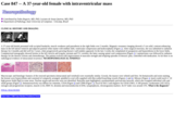
A 37-year-old female presented with occipital headache, muscle weakness and paresthesia in the right limbs over 3 months. Magnetic resonance imaging showed a 5 cm solid, contrast-enhancing mass in the left lateral ventricle and adjacent parietal white matter with midline shift, ventricular compression and hydrocephalus (Figure 1). After surgical resection, she was submitted to radiation therapy for 4 months and did well for 3 years, when progressively growing thoracic wall nodules appeared. In the last 2 weeks she complained of paraparesis and hypoesthesia in the lower limbs. Computerized tomography showed lesions in the left clavicle and scapula, sternum and T11 vertebra, the latter causing spinal cord compression (Figure 2). Laminectomy was followed by radiation therapy for the thoracic lesions and chemotherapy. The patient evolved with improvement of muscular strength and relapsing episodes of thoracic pain, controlled with medication. So far there is no radiological evidence of intracranial recurrence. NEUROPATHOLOGICAL FINDINGS
(This case study was added to OER Commons as one of a …
(This case study was added to OER Commons as one of a batch of over 700. It has relevant information which may include medical imagery, lab results, and history where relevant. A link to the final diagnosis can be found at the end of the case study for review. The first paragraph of the case study -- typically, but not always the clinical presentation -- is provided below.)
A 37-year-old man presented with a 2-month history of headache and balance impairment. He also complained of nausea and two episodes of vomiting a few days before admission. His past medical history was unremarkable. Neurological examination showed mild left sided ataxia.
(This case study was added to OER Commons as one of a …
(This case study was added to OER Commons as one of a batch of over 700. It has relevant information which may include medical imagery, lab results, and history where relevant. A link to the final diagnosis can be found at the end of the case study for review. The first paragraph of the case study -- typically, but not always the clinical presentation -- is provided below.)
37 year-old woman with no significant past medical history presents to a dermatologist for a "mole" on her left calf. The lesion has been present for approximately 6 months and besides the appearance has not been problematic. The patient has been otherwise asymptomatic. The patient's physical exam was within normal limits except for a 2 x 2cm erythematous, indurated nodule on her left medial calf. The patient's laboratory studies, including a complete blood count, a basic metabolic panel, erythrocyte sedimentation rate, rheumatoid factor, and antinuclear antibodies, were all within normal limits. A CT scan of the chest, abdomen, and pelvis was normal. The lesion was excised.
(This case study was added to OER Commons as one of a …
(This case study was added to OER Commons as one of a batch of over 700. It has relevant information which may include medical imagery, lab results, and history where relevant. A link to the final diagnosis can be found at the end of the case study for review. The first paragraph of the case study -- typically, but not always the clinical presentation -- is provided below.)
A female neonate was delivered at 38 weeks gestation to an 18 year-old, gravida 1, Native-American female whose antepartum course was notable for crystal methamphetamine and tobacco use early in pregnancy. Prenatal care had been initiated at 11 weeks of gestation, and an ultrasound performed at approximately 20 weeks revealed findings interpreted as a large posterior fossa cyst with mass effect. The cerebellum had not been well visualized, although the impression was that some cerebellar tissue was present. These features were overall felt to reflect a Dandy-Walker cyst. Additional ultrasonographic impressions included a probable porencephalic cyst on the right side, ventriculomegaly, possible agenesis of the corpus callosum, and overall significant absence of brain parenchyma in the right hemisphere. Serial follow-up ultrasound evaluations were performed at approximate 4 week intervals, demonstrating essentially the same intracranial ultrasonographic findings. Of note was that other organs showed appropriate growth progression. At delivery by Caesarian section, the posterior aspect of the neonate's scalp was noted to be covered by a thin, tense membrane, which ruptured during the procedure. Apgar scores were 7 and 9 at 1 minute and 5 minutes, respectively, and death occurred within hours of birth.
(This case study was added to OER Commons as one of a …
(This case study was added to OER Commons as one of a batch of over 700. It has relevant information which may include medical imagery, lab results, and history where relevant. A link to the final diagnosis can be found at the end of the case study for review. The first paragraph of the case study -- typically, but not always the clinical presentation -- is provided below.)
Our patient is a 38-year-old female with a past medical history significant for celiac sprue. She adheres strictly to a gluten-free diet. She is on no medications other than a multivitamin, and she has no family history of illnesses. She had her first child in 2001 (female) via normal spontaneous vaginal delivery and without complications.
(This case study was added to OER Commons as one of a …
(This case study was added to OER Commons as one of a batch of over 700. It has relevant information which may include medical imagery, lab results, and history where relevant. A link to the final diagnosis can be found at the end of the case study for review. The first paragraph of the case study -- typically, but not always the clinical presentation -- is provided below.)
A 38-year-old man, status 8 months post resection of his anaplastic oligodendrogioma of the right frontal lobe presented for a monitoring magnetic resonance imaging (MRI). The imaging studies revealed a slight interval increase of the minimally enhancing nodular mass consistent with the recurrent tumor (Figure 1). The patient received 6 cycles of temozolomide. Past medical history included frontal sinus infection and temporal mandibular joint dysfunction.
(This case study was added to OER Commons as one of a …
(This case study was added to OER Commons as one of a batch of over 700. It has relevant information which may include medical imagery, lab results, and history where relevant. A link to the final diagnosis can be found at the end of the case study for review. The first paragraph of the case study -- typically, but not always the clinical presentation -- is provided below.)
A 38 year-old female, known case of diabetes insipidus presented with a progressively deteriorating left sided weakness, headache and bilateral papilledema. Her lipid profile was normal. Magnetic resonance imaging revealed a large right-sided frontoparietal mass (8.5x5.8x4.7 cm) with smooth displacement of adjacent brain parenchyma and mild midline shift (Figures 1 and 2). A smaller (1.4x1.2x0.6 cm) yet similar lesion was identified on the right side of the falx cerebri. Radiologic diagnosis of meningioma was made. The patient underwent right frontoparietal craniotomy. A small amount of tissue was sent to pathology for intraoperative diagnosis followed by total resection of the large frontoparietal lesion. The lesion in the falx cerebri was not resected in view of its small size and location. No adjuvant therapy was administered.
(This case study was added to OER Commons as one of a …
(This case study was added to OER Commons as one of a batch of over 700. It has relevant information which may include medical imagery, lab results, and history where relevant. A link to the final diagnosis can be found at the end of the case study for review. The first paragraph of the case study -- typically, but not always the clinical presentation -- is provided below.)
This 39-year-old-woman had suffered from a moderate grade fever of unknown origin and a headache for 9 months. A previous hospital could not find the cause of her symptoms; therefore, she came to our hospital. At the first visit to our hospital, she had no signs of meningeal irritation and no palpable lymph nodes in the neck or supraclavicular regions. And then, she complained dysesthesia of her limbs and decreased visual acuity in the left eye. The laboratory data showed hypoproteinemia, hypoalbuminemia, hypochromic microcytic anemia, positive antinuclear antibody, and elevated serum levels of CRP (13.12 mg/dl), IgG (2080 mg/dl), IgA (622 mg/dl), and Interleukin-6 (IL-6) (73.2 pg/ml). CSF protein level was elevated (126 mg/dl), there was an oligoclonal band and IgG index was increased, but anti-aquaporin 4 (AQP4) antibody was negative. Cell count in CSF was normal. MRI detected an enhancing mass in the left cerebellar hemisphere (Figs.1A, 1B). In addition, there was a mildly enhancing lesion in the cervical spinal cord (Figs. 1C, 1D). But intraorbital region and optic nerve had no abnormalities. The patient had a suboccipital craniotomy for tumor resection. The tumor was connected to the choroid plexus at the foramen of Magendie without dural attachment, and was totally removed. The postoperative course was excellent, and no further fever appeared from the day after surgery. Immediately after the resection, dysesthesia of her limbs and visual disturbance in the left eye was also improved. The enhancing lesion in the cervical cord was vanished. Serum levels of CRP (0.1 mg/dl), IgG (1180 mg/dl) and IL-6 (0.8 pg/ml) were decreased and the anemia was improved after surgery.
(This case study was added to OER Commons as one of a …
(This case study was added to OER Commons as one of a batch of over 700. It has relevant information which may include medical imagery, lab results, and history where relevant. A link to the final diagnosis can be found at the end of the case study for review. The first paragraph of the case study -- typically, but not always the clinical presentation -- is provided below.)
A 39-year-old gentleman presented a single episode of hematuria and right flank pain of one month duration. Ultrasonography (USG) and CT of abdomen revealed a 7x5x5 cm right renal mass suggestive of renal cell carcinoma (RCC). While being investigated, he developed worsening headache and visual blurring. Positron emission tomography (PET) scan, CT (Figs. 1a, 1b) and post contrast MRI revealed a hyper-intense lesion in the right cerebellar hemisphere pushing the vermis to left and anteriorly (Figs. 1c, 1d). In view of raised intracranial pressure, he underwent gross total excision of the cerebellar lesion prior to nephrectomy.
(This case study was added to OER Commons as one of a …
(This case study was added to OER Commons as one of a batch of over 700. It has relevant information which may include medical imagery, lab results, and history where relevant. A link to the final diagnosis can be found at the end of the case study for review. The first paragraph of the case study -- typically, but not always the clinical presentation -- is provided below.)
A 39-year-old otherwise healthy woman was referred to our center in June 2002 due to progressive deterioration of vision in her left eye and visual field restriction of about two months' duration. Ever since she gave birth in 2000, she had been amenorrheic but still lactating. Neurological examination revealed bitemporal hemianopsia. Her endocrine function was normal.
(This case study was added to OER Commons as one of a …
(This case study was added to OER Commons as one of a batch of over 700. It has relevant information which may include medical imagery, lab results, and history where relevant. A link to the final diagnosis can be found at the end of the case study for review. The first paragraph of the case study -- typically, but not always the clinical presentation -- is provided below.)
The patient is a 39-year-old woman who had a left kidney tumor incidentally discovered during CT scan as part of a diagnostic workup for colonic diverticulosis. She had no personal or family history of significance, including no personal or family history of tuberous sclerosis complex (TSC), lymphangioleiomyomatosis, renal cyst, renal malignancy, or estrogen hormonal therapy. The CT scan demonstrated a 2.5-cm complex cystic mass in the upper pole of the left kidney with a 1-cm enhancing nodule in its wall, radiologically worrisome for cystic renal cell carcinoma. In view of this concern of malignancy, the patient elected to undergo laparoscopic left partial nephrectomy for definitive surgical treatment. The entire tumor was surgically resected with an excellent margin of 5-mm of normal parenchyma surrounding the entire cyst wall, and the tumor was confined to the kidney. She is alive with no evidence of recurrence or metastatic disease, 24 months postoperatively, and subsequent clinical follow-up with interval abdominal imaging studies is planned.
(This case study was added to OER Commons as one of a …
(This case study was added to OER Commons as one of a batch of over 700. It has relevant information which may include medical imagery, lab results, and history where relevant. A link to the final diagnosis can be found at the end of the case study for review. The first paragraph of the case study -- typically, but not always the clinical presentation -- is provided below.)
The patient is a 39-year-old white female presented with a 5 x 5 mm erythematous isolated papule on right forearm.
(This case study was added to OER Commons as one of a …
(This case study was added to OER Commons as one of a batch of over 700. It has relevant information which may include medical imagery, lab results, and history where relevant. A link to the final diagnosis can be found at the end of the case study for review. The first paragraph of the case study -- typically, but not always the clinical presentation -- is provided below.)
A 39 year-old-man with no significant past medical history presented with recurrent syncopal episodes, some of which were accompanied by nausea and vomiting. He also complained of recurrent headaches, gait problems and bilateral hand tremor. The physical exam confirmed the bilateral and symmetric intentional tremor and gait difficulties, without any motor or sensory deficits, including his cranial nerve examination. A brain MRI revealed a non-enhancing 2.4 x 1.7 cm mass within the right cerebellum; it was hypointense on T1 and hyperintense on T2 weighted sequences (Fig.1 and 2). Subtle leptomeningeal enhancement in the cortical sulci of both frontal lobes was suspicious for subarachnoid spread. Numerous T2 hyperintense lesions were also seen in the cervical and thoracic spine, the largest measuring 8 by 20 mm; these were similarly worrisome for CSF dissemination (Fig. 3 and 4). Providing further support for this notion, a cerebrospinal fluid specimen revealed scattered atypical small to medium sized cells with hyperchromatic nuclei, highly suspicious for malignant neoplasm. The patient subsequently underwent a subtotal resection of the cerebellar mass. Intra-operatively, the surgeon noted expanded cerebellar cortex with associated leptomeningeal coating or opacification suspicious for tumor involvement.
(This case study was added to OER Commons as one of a …
(This case study was added to OER Commons as one of a batch of over 700. It has relevant information which may include medical imagery, lab results, and history where relevant. A link to the final diagnosis can be found at the end of the case study for review. The first paragraph of the case study -- typically, but not always the clinical presentation -- is provided below.)
A 39-year-old man was admitted with recurrent fever, dizziness and headache of three months' duration. He related appetite loss and weight loss of 10 kg in the last three months. He had been diagnosed with HIV infection nine years before, but had never made use of highly active antiretroviral therapy (HAART). On physical examination, he was afebrile, confused, emaciated and presented oral candidiasis. There were no focal neurologic deficits or signs of meningeal irritation. Laboratory studies revealed a CD4 cell count of 101 cells/mm3, a viral load of 500,000 copies/ml, and serum sodium concentration of 145 mmol/liter. A blood test revealed increased levels of IgG for Toxoplasma gondii, with a negative IgM. A cerebrospinal fluid test revealed mild lymphocytic pleocytosis, elevated protein levels, and hypoglycorrhachia. No bacteria and fungi were identified with Gram, Ziehl-Neelsen, and India ink staining. At day 4, the serum sodium concentration increased to 155 mmol/liter. A computerized tomography of the head showed a slight edema and contrast enhancement in the hypothalamic area (Figure 1). At day 5, the patient complained of intense thirst and presented polyuria, hypotension, and elevated levels of serum urea and creatinine. The serum sodium concentration remained high (164 mmol/liter), even after hypotonic fluid infusion. Urine analysis revealed low osmolality, low sodium concentration, and low density. A diagnosis of probable central diabetes insipidus and acute renal failure due to dehydration was established and treatment with desmopressin by nasal spray was initiated. At day 7, the patient developed pneumonia and his general condition worsened. He was transferred to the Intensive Treatment Unit and maintained under the same antibiotic regime. He died two days later (nine days after admission). A post-mortem brain MRI showed symmetric hyperintensities at the floor of the third ventricle in the hypothalamus on axial T2-weighted images (Figure 2).
(This case study was added to OER Commons as one of a …
(This case study was added to OER Commons as one of a batch of over 700. It has relevant information which may include medical imagery, lab results, and history where relevant. A link to the final diagnosis can be found at the end of the case study for review. The first paragraph of the case study -- typically, but not always the clinical presentation -- is provided below.)
A 39-year-old man presented to a small local hospital in northern Saskatchewan following new-onset seizure activity. A single seizure began focally in the left arm and subsequently became secondarily generalized. He was observed in hospital for two days, during which there were no further seizures. He was released after arrangements had been made for him to follow-up with a neurologist, an appointment which he did not keep. Two months later he presented to our institution after having had three similar seizures in short succession. On examination, he was alert and oriented, but drowsy. There was a mild dysphasia, and a partial right-sided visual field defect. Physical examination was otherwise relatively unremarkable. There was no history of headache, nausea, vomiting, weakness, altered sensation, or fever. There were no significant past medical or surgical histories. The patient occasionally smoked cigarettes, had previously abused alcohol, and had previously been exposed to tuberculosis.
(This case study was added to OER Commons as one of a …
(This case study was added to OER Commons as one of a batch of over 700. It has relevant information which may include medical imagery, lab results, and history where relevant. A link to the final diagnosis can be found at the end of the case study for review. The first paragraph of the case study -- typically, but not always the clinical presentation -- is provided below.)
A 3-month-old boy was referred to our hospital because of slowly progressing weakness, which commenced a month earlier. He was delivered normally from an uneventful pregnancy from nonconsanguineous parents. A neonatal screening test had revealed mildly increased serum carnitine, which was normal in a repeated test. He showed generalized hypotonia with an absence of deep tendon reflexes and a lagged head on neurological examination. During the admission, respiratory muscle weakness progressed and he became dependent on mechanical ventilation with tracheal fenestration. Laboratory tests were unremarkable except for increased aspartate aminotransferase (154 IU/L), alanine aminotransferase (68 IU/L), and creatinine kinase (478 U/L) levels. Multiplex ligation-dependent probe amplifications for SMN1/SMN2, the causative gene for spinal muscular atrophy (SMA) type I, revealed no deletion or duplication. He had additional tests, including metabolic screening, brain MRI, and muscle biopsy. Brain MRI was unremarkable for his age, whereas metabolic screening revealed elevated glutaric acid and ethylmalonic acid in his urine, and elevated plasma C14, C14:1, and C16:1 carnitines.
(This case study was added to OER Commons as one of a …
(This case study was added to OER Commons as one of a batch of over 700. It has relevant information which may include medical imagery, lab results, and history where relevant. A link to the final diagnosis can be found at the end of the case study for review. The first paragraph of the case study -- typically, but not always the clinical presentation -- is provided below.)
This 3-month-old girl was referred to the Neurology Clinic for hypotonia. The child was born at 36 weeks to a 22-year-old G1P0 mother with an uncomplicated pregnancy. The neonatal period was uneventful, however, in the first few months, the pediatrician noted that the child seemed hypotonic.
No restrictions on your remixing, redistributing, or making derivative works. Give credit to the author, as required.
Your remixing, redistributing, or making derivatives works comes with some restrictions, including how it is shared.
Your redistributing comes with some restrictions. Do not remix or make derivative works.
Most restrictive license type. Prohibits most uses, sharing, and any changes.
Copyrighted materials, available under Fair Use and the TEACH Act for US-based educators, or other custom arrangements. Go to the resource provider to see their individual restrictions.