(This case study was added to OER Commons as one of a …
(This case study was added to OER Commons as one of a batch of over 700. It has relevant information which may include medical imagery, lab results, and history where relevant. A link to the final diagnosis can be found at the end of the case study for review. The first paragraph of the case study -- typically, but not always the clinical presentation -- is provided below.)
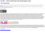
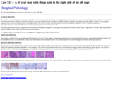
A 3-year-old boy was referred for pre-surgical evaluation due to drug-resistant complex partial seizures. The patient was born to unrelated healthy parents and had developed normally. There was no family history of any central nervous system (CNS) disease. His seizures started at the age of 4 months and were characterized by daily, brief (30-60 seconds) episodes of staring, gestural automatisms, and tachycardia, followed by somnolence or headache. From age 14 months the boy started suffering also from frequent left focal motor seizures often resulting in secondarily generalization. At our observation, seizures still occurred daily despite treatment with several antiepileptic drugs. In addition, his parents had also noted initial worsening of cognitive function. Dermatologic evaluation revealed a large congenital nevus on the scalp (Fig 1) and two small black pigmented nevi on gluteus and abdomen. Electroencephalographic recordings showed frequent slow and sharp waves over the right temporal region (Figs 2 and 3). Brain MRI showed a focal lesion in the right uncus. The lesion was hyperintense on T1-weighted (Figs 4 and 5) and hypointense on T2-weighted images (Fig 6) with no gadolinium enhancement. No mass effect or surrounding edema was evident. A right anterior temporal lobectomy and hippocampectomy was performed (Figs 7, 8 and 9). Intraoperative electrocorticography before resection revealed active spikes around the lesion. The postoperative course was uneventful. After surgery, the boy remained seizure-free at the 15-months follow-up.
(This case study was added to OER Commons as one of a …
(This case study was added to OER Commons as one of a batch of over 700. It has relevant information which may include medical imagery, lab results, and history where relevant. A link to the final diagnosis can be found at the end of the case study for review. The first paragraph of the case study -- typically, but not always the clinical presentation -- is provided below.)
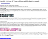
A 3-year-old girl with a history of mild motor delay, presented with a change in mental status, gait difficulty, nausea and vomiting. The patient was in her usual state of health until 9 months prior to presentation, when she was found to have abnormal adduction of the left eye. Over the next several months, she developed gait difficulties and behavioral changes. On the day of admission, she developed confusion, severe gait difficulties, nausea and vomiting. On physical examination, she had right homonymous hemianopsia, right hemiparesis, and increased deep tendon reflexes of her right Achilles tendon. She subsequently became unresponsive. She was intubated and given mannitol and steroids. An external ventricular drain was placed. Neuroimaging showed a minimally heterogeneous enhancing tumor measuring 8 x 8 x 9 cm involving the left parietal region and, partly filling the left lateral ventricle, with accompanying hydrocephalus, midline shift and uncal herniation (Figures 1, 2, 3). Gross total resection of the tumor was confirmed by post-operative imaging. Staging evaluation revealed no metastasis. The patient was discharged one week later.
(This case study was added to OER Commons as one of a …
(This case study was added to OER Commons as one of a batch of over 700. It has relevant information which may include medical imagery, lab results, and history where relevant. A link to the final diagnosis can be found at the end of the case study for review. The first paragraph of the case study -- typically, but not always the clinical presentation -- is provided below.)
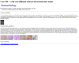
The patient is a 3 year old male who initially presented with seizures at age 18 months and was diagnosed with a Chiari malformation; this was successfully surgically repaired. During the pre-operative assessment, however, he was found to have isolated hepatomegaly. Since that time, his parents have noted increasing fatigue, leg pain, intermittent abdominal pain, and a possible two pound weight loss. Repeated blood sugar measurements revealed glucose levels in the 30s and 40s in the morning and early afternoon. He has no history of jaundice, or pruritus. There is no family history of similar symptoms (see Figure 1).
(This case study was added to OER Commons as one of a …
(This case study was added to OER Commons as one of a batch of over 700. It has relevant information which may include medical imagery, lab results, and history where relevant. A link to the final diagnosis can be found at the end of the case study for review. The first paragraph of the case study -- typically, but not always the clinical presentation -- is provided below.)
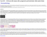
A 40 year old female presented to the neurosurgery department with a history of new onset, severe, pressure-like headaches. MRI (Fig. 1) revealed multiple mass lesions in the spinal cord, at C4-6 and T3 with infiltration of the neuroforamina. An epidural component was associated with spinal cord compression. In addition, there were lesions in the hip and shoulder, liver and small intestine, as well as right parietal and frontal, dural- based lesions, 3x3x4 and 1.5x1x1cm. Subsequent surgical removal of the dural tumors was complete. The postoperative course was uneventful and the patient described relief from the headaches. The intracranial tumors recurred at 1 and 2 years after the initial surgery.
(This case study was added to OER Commons as one of a …
(This case study was added to OER Commons as one of a batch of over 700. It has relevant information which may include medical imagery, lab results, and history where relevant. A link to the final diagnosis can be found at the end of the case study for review. The first paragraph of the case study -- typically, but not always the clinical presentation -- is provided below.)
A 40-year-old right-handed female first noticed 6 months ago headache and dizziness. One month later, she complained of repeated vomiting and frequent headache. She was hospitalized at another medical facility. MRI examinations revealed multiple contrast enhanced lesions over the surface of cerebral hemispheres (Figure 1), cerebellum, brainstem (Figure 2) and cervical spinal cord (Figure 3). Laboratory examinations showed no abnormality in the white blood cell (WBC) count, C-reactive protein (CRP), or the tumor markers such as CEA, CA19-9 and SCC. Lumbar punctures revealed an increased opening pressure of 280 mm H2O. Analysis of the cerebrospinal fluid (CSF) disclosed mildly elevated cell counts (22 cells/mm3; 86% mononuclear, 14% polymorphonuclear), a severely decreased concentration of glucose (4 mg/dl) and elevated level of protein (446 mg/dl). A polymerase chain reaction (PCR) test for tuberculosis DNA in the CSF was reported to be normal, and negative result of tuberculosis cultures. No clinical improvement was observed despite a trial of steroid therapy. Because these studies did not reveal a diagnosis and she became tetraparetic, an open biopsy was performed through a left temporal craniotomy 4 months after the onset of initial symptom. At surgery, milky opacification of the leptomeninges was observed at the convexity of the brain and they were biopsied with adjacent brain tissue. Five months after the onset of the disease, she was transferred to our hospital. At this time, she showed tetraparesis (Manual Mascle Test (MMT): Upper extremity=3/5, Lower extremity=1/5), dysphagia, right hearing disturbance, left oculomotor paralysis, sensory disorders of the bilateral lower extremities, and bladder and rectal disturbances. The Karnofsky Performance Scale (KPS) was 40.
(This case study was added to OER Commons as one of a …
(This case study was added to OER Commons as one of a batch of over 700. It has relevant information which may include medical imagery, lab results, and history where relevant. A link to the final diagnosis can be found at the end of the case study for review. The first paragraph of the case study -- typically, but not always the clinical presentation -- is provided below.)
A 40-year-old woman presented to the Neurosurgery Department with sudden onset of headache and altered sensorium for 2 days. On evaluation in the emergency department, she was drowsy, disoriented with Glasgow coma scale of 13/15 and right hemiparesis grade 3/5, with no cranial nerve deficits. She was earlier diagnosed to have diffuse osteomalacia three years back and was on treatment for the same. Her serum phosphate levels (1.5mg/dl) were low, calcium levels (9.2mg/dl) were normal and vitamin D levels (40nmol/l) were reduced. Cause for the same could not be ascertained despite several investigations.
(This case study was added to OER Commons as one of a …
(This case study was added to OER Commons as one of a batch of over 700. It has relevant information which may include medical imagery, lab results, and history where relevant. A link to the final diagnosis can be found at the end of the case study for review. The first paragraph of the case study -- typically, but not always the clinical presentation -- is provided below.)
A 40-year-old male with a one year clinical history of vertigo and intermittent headaches was admitted for evaluation at a tertiary university hospital. Magnetic Resonance Image (MRI) showed a heterogenously enhancing mass with cystic and calcific components in the anterior horn of the left lateral ventricle measuring 2.5 x 2.2 x 1.6 cm (Fig. 1). The patient underwent partial resection of the intraventricular tumor through a left frontal craniotomy with transfrontal approach to the left ventricle. The patient was discharged four days later and has done well since that time with no neurological deficits.
(This case study was added to OER Commons as one of a …
(This case study was added to OER Commons as one of a batch of over 700. It has relevant information which may include medical imagery, lab results, and history where relevant. A link to the final diagnosis can be found at the end of the case study for review. The first paragraph of the case study -- typically, but not always the clinical presentation -- is provided below.)
A 40-year-old female presented with blurred vision and diplopia, followed by slowly progressive left-sided motor and sensory disturbances. She also suffered from memory loss and was had mild spatial and temporal disorientation. A T2 weighted MRI showed a large area of high signal intensity in the periventricular white matter of the right more than the left occipital region and the corpus callosum, without enhancement on T1 weighted images after gadolinium administration and without mass effect. A stereotactic biopsy of the intracerebral lesion showed blast-like neoplastic cells within a mononuclear infiltrate. No diagnosis could be made based on morphology and immunohistochemistry using a large series of markers. However, based on positive OCT3/4 nuclear staining, the tumor was diagnosed as a germinoma (seminoma of the brain). The patient was treated accordingly and her condition improved, although focal deficits remained.
(This case study was added to OER Commons as one of a …
(This case study was added to OER Commons as one of a batch of over 700. It has relevant information which may include medical imagery, lab results, and history where relevant. A link to the final diagnosis can be found at the end of the case study for review. The first paragraph of the case study -- typically, but not always the clinical presentation -- is provided below.)
A 40-year-old female presented with new-onset seizures 4 months ago and with a menstrual disorder for the past 2 months. She was admitted to our hospital for evaluation. Physical examination was non-specific. MRI of the brain revealed a well-delineated homogeneously contrast-enhancing mass in the posterior lobe of the hypophysis. No abnormal signal was found in brain parenchyma. Serum levels for LH, FSH and prolactin were within normal limits as were progesterone and estrogen. MRI demonstrated a sellar mass measuring about 3.6×3.5 mm in size. The lesion was generally isointense to gray matter on T1-weighted images (Figure 1) and isointense to slightly hyperintense on T2-weighted images (Figure 2). The patient was diagnosed with a presumed pituitary microadenoma and epilepsy, and an endoscopic transphenoidal resection was performed.
(This case study was added to OER Commons as one of a …
(This case study was added to OER Commons as one of a batch of over 700. It has relevant information which may include medical imagery, lab results, and history where relevant. A link to the final diagnosis can be found at the end of the case study for review. The first paragraph of the case study -- typically, but not always the clinical presentation -- is provided below.)
A 41 -year-old male patient administered to hospital with the complaint of medically intractable complex partial seizures since 3 years of age. The frequency of focal seizures despite multi-medication treatment was 4-5 /month. Brain MR imaging revealed a non-enhancing cortical lesion with subcortical involvement in the left parietal lobe. Imaging characteristics of the lesion on T1-W was peculiar in that both hypo- and isointensity was present with accompanying peripheral hyperintensity (Fig. 1). The lesion was hyperintense with microcysts on FLAIR (Fig. 2). Following lobectomy the patient remained seizure free and showed no evidence of tumor recurrence in the 48-months follow-up period.
(This case study was added to OER Commons as one of a …
(This case study was added to OER Commons as one of a batch of over 700. It has relevant information which may include medical imagery, lab results, and history where relevant. A link to the final diagnosis can be found at the end of the case study for review. The first paragraph of the case study -- typically, but not always the clinical presentation -- is provided below.)
A 41-year-old previously healthy Japanese female presented with a 2-month-history of left occipital headache and dizziness. She had instability in standing on her left foot. A contrast-enhanced T1-weighted MR scan showed a high signal, 4.6 × 3.2 × 3.5 cm extra-axial mass containing a cyst located in the left posterior cranial fossa (Fig. 1a). It attached to the dura mater along the occipital bone and the petrous bone, a part of which was extending into the sigmoid sinus. Catheter angiography illustrated a hypervascular tumor fed by the middle meningeal artery and the ascending pharyngeal artery. The sigmoid sinus was occluded by the tumor invasion.
(This case study was added to OER Commons as one of a …
(This case study was added to OER Commons as one of a batch of over 700. It has relevant information which may include medical imagery, lab results, and history where relevant. A link to the final diagnosis can be found at the end of the case study for review. The first paragraph of the case study -- typically, but not always the clinical presentation -- is provided below.)
The patient is a 41 year old gentleman who presented with a 2 week history of right sided "sharp and stabbing" rib pain without history of trauma or injury. The patient stated he felt a "snap" with a burst of pain in the right costal area, followed by inability to walk and could not breathe secondary to pain. His weight and appetite were stable prior to the incident. A chest CT scan revealed expansion of the cortex and medullary cavity of the right posterior 6th rib and a fracture with associated lytic focus in the posterior right 9th rib. The CT scan was interpreted as "hyperplasia/dysplasia" of the right 6th posterior rib and a pathologic fracture of the 9th posterior rib.
(This case study was added to OER Commons as one of a …
(This case study was added to OER Commons as one of a batch of over 700. It has relevant information which may include medical imagery, lab results, and history where relevant. A link to the final diagnosis can be found at the end of the case study for review. The first paragraph of the case study -- typically, but not always the clinical presentation -- is provided below.)
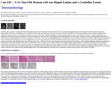
In 1995, at the age of 29, a woman presented with headache and visual disturbances. She was found to have a capillary hemangioblastoma of the brainstem, which was then resected and pathologically confirmed. The diagnosis of von Hippel-Lindau disease was suspected at this time. One year later, she presented with worsening headache, nausea, vertigo, photophobia, and episodes of unresponsiveness. CT and MRI revealed a 4 cm mass in the right cerebellar hemisphere with effacement and displacement of the fourth ventricle. A VP shunt was placed semi-emergently, and two days later she underwent a second craniotomy for tumor resection. The diagnosis of hemangioblastoma was once again confirmed. In 2004, follow-up MRI of the brain and spine revealed multiple brainstem and cervical and thoracic cord lesions consistent with hemangioblastomas. An abdominal scan showed cystic kidneys with bilateral enhancing heterogeneous renal masses, suspicious for malignancy, as well as multiple pancreatic cysts and a paraaortic nodule of possible left adrenal origin; however a bone scan was normal. It is unclear whether or not the patient was treated for the presumed renal cell carcinoma at this time. In 2007, the then 41-year-old patient presented with a three-day history of falls and disequilibrium, progressive quadriparesis and difficulty swallowing. An MRI of the brain was performed. Axial T2 (Figure 1) and contrast enhanced T1-weighted (Figure 2) images showed a partially cystic (thick arrow) enhancing cerebellar mass, dorsal to the fourth ventricle. Extensive T2 hyperintensity was present in the pons, medulla (thin arrows) and middle cerebellar peduncles. Sagittal unenhanced (Figure 3) and contrast enhanced (Figure 4) T1-weighted images showed a markedly enhancing (black arrowhead) solid mass inferior to the fourth ventricle. Nodular leptomeningeal enhancement on the anterior surface of the pons was consistent with a leptomeningeal tumor. The patient underwent a posterior fossa craniotomy with gross total resection of the mass in April 2007.
(This case study was added to OER Commons as one of a …
(This case study was added to OER Commons as one of a batch of over 700. It has relevant information which may include medical imagery, lab results, and history where relevant. A link to the final diagnosis can be found at the end of the case study for review. The first paragraph of the case study -- typically, but not always the clinical presentation -- is provided below.)
A previously healthy 42-year-old man presented with two weeks of confusion, lethargy, headache, and malaise. He was diagnosed with AIDS three months prior (CD4 count 198 cells/microliter; viral load 87,100 copies/mL) and was on trimethoprim/sulfamethoxazole prophylaxis but had not started highly active antiretroviral therapy. Neurologic exam was notable for a sleepy man lacking orientation to location or date, perseveration on simple phrases and overall paucity of speech, and generalized weakness. He could not stand unassisted. Reflexes were 3+ throughout.
(This case study was added to OER Commons as one of a …
(This case study was added to OER Commons as one of a batch of over 700. It has relevant information which may include medical imagery, lab results, and history where relevant. A link to the final diagnosis can be found at the end of the case study for review. The first paragraph of the case study -- typically, but not always the clinical presentation -- is provided below.)
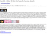
A 42 year old man with history of asthma, sinusitis and GERD presented to the critical care unit with a 6 day history of worsening headache, nausea, and vomiting. He reported that the symptoms began with a "popping" sensation after a bout of coughing and fever, although he was afebrile on admission. Neurological exam revealed that he was sleepy, but easily arousable and oriented x 3 with a non-focal motor exam. CT scan showed a large right temporal-occipital hyperdense lesion with surrounding edema suggestive of an acute/subacute hemorrhage and a 4-5mm associated midline shift with no evidence of hydrocephalus. On day 2 he developed a 4/5 left sided hemiparesis, dysarthria, and left hemifacial weakness. MRI of brain demonstrated a large right temporal-occipital intraparenchymal hemorrhage, subtle leptomeningeal enhancement, an increased T2 signal of the brainstem especially within the right pons, and a partial thrombosis of the right transverse and sigmoid sinuses. There was no evidence of underlying tumor or vascular malformations. He developed a worsening of the left hemiparesis (2/5) and was started on heparin anticoagulation for a suspected venous infarct, secondary to the venous sinus thrombosis. On hospital day 4 he was noted to be more alert with improvement in his speech. Repeat MRI showed recanalization of the transverse sinus and stable abnormalities in the brainstem and right temporal-occipital lobes. He remained awake, alert, and oriented with some improvement in his hemiparesis. On day 8 he had a sudden neurological decline, became hemodynamically unresponsive and ultimately expired despite aggressive medical management.
(This case study was added to OER Commons as one of a …
(This case study was added to OER Commons as one of a batch of over 700. It has relevant information which may include medical imagery, lab results, and history where relevant. A link to the final diagnosis can be found at the end of the case study for review. The first paragraph of the case study -- typically, but not always the clinical presentation -- is provided below.)
A 42 year old Caucasian male patient with known Hashimoto's thyroiditis presented the first time in 2010 with increased thirst and nocturia. An MRI of the pituitary gland in 2010 showed an enlarged pituitary stalk up to the infundibulum. Endocrinologically, there were normal levels for ACTH, LH, FSH, TSH, PRL, STH as well as for cortisol, testosterone and IGF-1. The clinical diagnosis of diabetes insipidus due to an autoinflammatory hypophysitis with a normal functioning anterior pituitary was made after excluding other reasons (e.g. sarcoidosis). A symptomatic treatment with desmopressin was initiated and regular follow-ups were held.
(This case study was added to OER Commons as one of a …
(This case study was added to OER Commons as one of a batch of over 700. It has relevant information which may include medical imagery, lab results, and history where relevant. A link to the final diagnosis can be found at the end of the case study for review. The first paragraph of the case study -- typically, but not always the clinical presentation -- is provided below.)
A 42-year-old male suffered a witnessed, new-onset generalized tonic-clonic seizure while driving a car, which lead to an accident. In addition to minor injuries, trauma work-up revealed a 3.6 x 2.6 x 3 cm dural-based mass compressing the right middle temporal gyrus. The lesion appeared well-circumscribed, oval in shape, showed moderate patchy contrast enhancement (Fig. 1). In addition, fine T2- and T1-hyperintense rimming was noted, accompanied by mild perifocal edema, and interestingly, homogenous hypointensity on T2-weighted images (Fig. 2). Evidence of calcification was absent on cranial computed tomography (not shown). Calcification was seen within a small (0.7x2.5x1 cm), fusiform lesion with homogenous contrast enhancement localized in the frontal falx, consistent with a meningioma (Fig. 3). The patient subsequently underwent right temporal osteoplastic craniotomy for the temporal lobe lesion. Intraoperatively, the lesion was adherent to the dura and resembled a meningioma with typical meningeal blood supply. After circular incision and coagulation of the dura, the tumor showed a glassy-whitish appearance. The rubber-like, cartilaginous consistency made it impossible to debulk the central components, despite highest volume of the ultrasonic surgical aspirator. Fortunately, the arachnoidal layer was intact throughout and adhesions were minimal. The core of the tumor could be partially resected using a regular scalpel and then the lesion was shelled-out en bloc (Fig. 4). Finally, the surgical result resembled a Simpson grade I resection of a meningioma.
(This case study was added to OER Commons as one of a …
(This case study was added to OER Commons as one of a batch of over 700. It has relevant information which may include medical imagery, lab results, and history where relevant. A link to the final diagnosis can be found at the end of the case study for review. The first paragraph of the case study -- typically, but not always the clinical presentation -- is provided below.)
A 42-year-old white male presented with sudden onset of suprapubic and pelvic discomfort associated with gross hematuria. His vitals were stable on admission. A cystogram demonstrated a bladder of normal size and contour with no intravesical filling defect. Computed tomography (CT) revealed thickened bladder wall with possible infiltrating hematoma and obstruction of the right ureter with hydronephrosis.
(This case study was added to OER Commons as one of a …
(This case study was added to OER Commons as one of a batch of over 700. It has relevant information which may include medical imagery, lab results, and history where relevant. A link to the final diagnosis can be found at the end of the case study for review. The first paragraph of the case study -- typically, but not always the clinical presentation -- is provided below.)
A 42-year-old man was admitted to the neurosurgery department because of paraparesis and sensory deficits of both feet. The CT scan and MRI examination revealed a solitary intramedullary lesion bulging dorsally from the thoracic spine (T4 level) (Figure 1). Spinal angiography revealed the dense vascularity of the lesion, the presence of feeding and draining vessels, as well as intra-lesional shunting. A gross total resection was performed.
No restrictions on your remixing, redistributing, or making derivative works. Give credit to the author, as required.
Your remixing, redistributing, or making derivatives works comes with some restrictions, including how it is shared.
Your redistributing comes with some restrictions. Do not remix or make derivative works.
Most restrictive license type. Prohibits most uses, sharing, and any changes.
Copyrighted materials, available under Fair Use and the TEACH Act for US-based educators, or other custom arrangements. Go to the resource provider to see their individual restrictions.