(This case study was added to OER Commons as one of a …
(This case study was added to OER Commons as one of a batch of over 700. It has relevant information which may include medical imagery, lab results, and history where relevant. A link to the final diagnosis can be found at the end of the case study for review. The first paragraph of the case study -- typically, but not always the clinical presentation -- is provided below.)
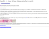
A 42-year-old man presented with three months of progressive proximal weakness, a thirty pound weight loss, and worsening dysphagia for solids. He had been hospitalized two months earlier for respiratory failure requiring intubation and five days of mechanical ventilation. Arterial blood gas on admission revealed a pH of 7.26; pCO2 58.8 torr; bicarbonate 26.2; and pO2 of 57.1 torr. A primary pulmonary etiology was not found, and he improved. During that hospitalization, an electromyogram of the upper extremities was normal. There were chronic neurogenic changes in the lower extremities. Low amplitude, short duration motor unit potentials suggestive of myopathy were noted in the cervical and thoracic paraspinal muscles.
(This case study was added to OER Commons as one of a …
(This case study was added to OER Commons as one of a batch of over 700. It has relevant information which may include medical imagery, lab results, and history where relevant. A link to the final diagnosis can be found at the end of the case study for review. The first paragraph of the case study -- typically, but not always the clinical presentation -- is provided below.)
A 42-year-old previously healthy male with a past medical history significant only for hyperlipidemia presented with weakness, fatigue and edema of the face and extremities. Initial laboratory studies demonstrated anemia (9.8 mg/dL), renal insufficiency (creatinine 2.3 mg/dL), a weak positive ANA of 1:80 with nucleolar pattern, elevated rheumatoid factor, and negative hepatitis B and C serologies. Urine sediment contained a small number of erythrocytes and white blood cells, and granular and hyaline casts. Because of the patient's clinical picture, including moderate anemia, a serologic evaluation for parvovirus B19 was performed, which was positive for both IgG and IgM antibodies. Additionally, because of renal failure, a renal biopsy was performed.
(This case study was added to OER Commons as one of a …
(This case study was added to OER Commons as one of a batch of over 700. It has relevant information which may include medical imagery, lab results, and history where relevant. A link to the final diagnosis can be found at the end of the case study for review. The first paragraph of the case study -- typically, but not always the clinical presentation -- is provided below.)
A 42 year old previously healthy woman presented with an enlarging soft tissue mass in the popliteal fossa of the left posterior knee. The mass had been noticed 4 months prior and recently began to interfere with flexion of the joint. No preceding trauma was noted.
(This case study was added to OER Commons as one of a …
(This case study was added to OER Commons as one of a batch of over 700. It has relevant information which may include medical imagery, lab results, and history where relevant. A link to the final diagnosis can be found at the end of the case study for review. The first paragraph of the case study -- typically, but not always the clinical presentation -- is provided below.)
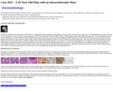
A 43-year-old woman presented with a six month history of progressive nocturnal nausea and vomiting, resulting in 30 kilograms of weight loss. Subsequently, she developed gradual cognitive decline and a cerebellar syndrome with gait ataxia and nystagmus. MRI showed multifocal lesions localized periventricularly, parenchymal and in the pons on contrast-enhanced T1-weighted images (Figure 1). Examination of cerebrospinal fluid (CSF), whole body positron emission tomography computerized tomography (PET-CT) scan and bone marrow biopsy did not show extra cranial involvement. The differential diagnosis consisted of metastatic melanoma, lymphoma, multiple sclerosis, meningoencephalitis or a granulomatous infection. Stereotactic biopsy of the lesions was performed.
(This case study was added to OER Commons as one of a …
(This case study was added to OER Commons as one of a batch of over 700. It has relevant information which may include medical imagery, lab results, and history where relevant. A link to the final diagnosis can be found at the end of the case study for review. The first paragraph of the case study -- typically, but not always the clinical presentation -- is provided below.)
A previously healthy 43-year-old woman first suffered from headache with dizziness, vomiting, and nausea beginning 5 months before hospital admission. CT scans showed subarachnoid hemorrhage involving the right temporal and occipital lobes, while MRI revealed abnormal signals in the left thalamus and right temporal-occipital lobes. Five months later she suffered from headache, transient unconsciousness and aggravated left upper-limb paralysis. This time, neurological examinations revealed a decrease in power and deep tendon reflexes of the left upper limb. The activated partial thromboplastin time was notably prolonged at 38.8s, while the prothrombin time was normal. Cerebrospinal fluid (CSF) contained 100 erythrocytes /mm3. Neuroimaging revealed infarcts and hemorrhages in bilateral frontal lobes (Fig. 1a) and slit-like complex softening of the right temporal-occipital lobes (Fig. 1b). MRI showed diffuse enhancement in the dural mater and sulci of bilateral frontal-parietal lobes and tentorium (Fig. 1b).MR venography disclosed occlusions in the right transverse and sigmoid sinus, suggesting thrombosis (Fig. 1c). Five days after admission, the patient died and a restricted cranial autopsy was performed.
(This case study was added to OER Commons as one of a …
(This case study was added to OER Commons as one of a batch of over 700. It has relevant information which may include medical imagery, lab results, and history where relevant. A link to the final diagnosis can be found at the end of the case study for review. The first paragraph of the case study -- typically, but not always the clinical presentation -- is provided below.)
A 43-year-old gentleman presented with swelling over scalp for past four months which was initially painless, hard and progressively increasing in size. It became painful in last one month, which was dull aching, intermittent and relieved by medication. There was no history of seizures, weakness, loss of consciousness, trauma, blurring of vision or vomiting. On examination, the vitals were stable with Glasgow coma scale of E4-V5-M6. Bilateral pupils were of normal size with normal reaction. There were no sensory or motor deficits and cerebellar signs were negative. Contrast enhancing MRI showed bone destructive lesion over parietal region. Axial CT showed a lytic lesion of the calvaria in the midline (Fig. 1a). Some expansion of bone with sclerosis was noted. Sagittal and coronal MRI revealed a midline parietal calvarial lesion, indenting the intact underlying dural structures and expanding the overlying galea (Figs. 1b, 1c). With a preoperative diagnosis of midline fronto-parietal calvarial tumor, a midline fronto-parietal craniotomy was performed and near total excision was achieved. Intraoperatively, a firm to hard pinkish tumor was visualized in the calvarium which was adherent to dura and infiltrating through the calvarial bone. Entire tumor with surrounding 1 cm of bony margin was excised. Post-operative period was uneventful.
(This case study was added to OER Commons as one of a …
(This case study was added to OER Commons as one of a batch of over 700. It has relevant information which may include medical imagery, lab results, and history where relevant. A link to the final diagnosis can be found at the end of the case study for review. The first paragraph of the case study -- typically, but not always the clinical presentation -- is provided below.)
A 43 year old man presented 22 years ago with proptosis and reduced visual acuity of the right eye. He was investigated and found to have an orbital tumor which was subsequently biopsied but not resected. He currently presented with decreased visual acuity and impaired right-sided gaze. On imaging, there was a right retro-orbital tumor for which an excision was performed. Intraoperatively, a well-encapsulated tumor within the cystic component containing clear fluid was noted and the relation of this tumor to the nerve could not be clearly ascertained. On histology, the excised specimen revealed a cellular neoplasm consisting of sweeping fascicles of spindle cells with storiform appearance in some areas. Focal ill defined, Verocay body-like palisades were identified. Scattered blood vessels with hyalinised wall were noted. There was no necrosis, significant atypia or mitotic activity. The tumor showed strong staining for S-100 and focal staining for glial fibrillary acidic protein (GFAP) on immunohistochemistry and ultrastructurally, numerous intertwining cell processes were discerned. The findings corroborated a diagnosis of retrobulbar cellular schwannoma, the retrobulbar region being an uncommon location site for this tumor. Important differential diagnoses to be considered include meningioma, glial tumor in view of positive staining for GFAP in cellular schwannoma and malignant peripheral nerve sheath tumor (MPNST) in view of the increased cellularity. While metastases and death have not been described in cellular schwannoma, incompletely resected cellular schwannoma has presented with recurrence. In such a situation, mitotic count significantly correlates with the incidence of tumor recurrence and close follow up is recommended.
(This case study was added to OER Commons as one of a …
(This case study was added to OER Commons as one of a batch of over 700. It has relevant information which may include medical imagery, lab results, and history where relevant. A link to the final diagnosis can be found at the end of the case study for review. The first paragraph of the case study -- typically, but not always the clinical presentation -- is provided below.)
The patient is a 43 year old morbidly obese man with chronic back pain, progressively increasing in severity and unresponsive to medical management. A magnetic resonance scan performed at an outside institution revealed a spinal mass at the L4-L5 vertebral level. The patient was referred to UPMC and underwent a laminectomy of the L4 and L5 vertebrae, and upper portion of the sacrum. Intraoperatively, the lesion was described as a "huge sausage-shaped mass approximately 3.5 cm to 4 cm long with all of the nerve roots peripherally draped over the mass." The mass was arising from the filum terminale and the surface appeared to be well-encapsulated. Complete resection of the tumor was achieved. The post-operative diagnosis was recorded as "intradural spinal cord tumor (ependymoma) L4, L5, S1."
(This case study was added to OER Commons as one of a …
(This case study was added to OER Commons as one of a batch of over 700. It has relevant information which may include medical imagery, lab results, and history where relevant. A link to the final diagnosis can be found at the end of the case study for review. The first paragraph of the case study -- typically, but not always the clinical presentation -- is provided below.)
A 43-year-old woman was admitted with headache over a 3 week period. The clinical examination was completely unremarkable. The past medical history included breast cancer three years before presentation with a contralateral relapse another two years later. Treatment consisted of surgical resection, radiotherapy and post-operative chemotherapy with 4 cycles of epirubicine and cyclophosphamide. The relapse was treated by resection, radiotherapy and anti-estrogen therapy. Two years before current admission, acute myeloid leukemia FAB M4 Eo was diagnosed and treated with induction and consolidation chemotherapy according to the German AMLCG protocol with TAD/HAM double-induction and TAD consolidation chemotherapy followed by 4 weekly alternating maintenance chemotherapy.
(This case study was added to OER Commons as one of a …
(This case study was added to OER Commons as one of a batch of over 700. It has relevant information which may include medical imagery, lab results, and history where relevant. A link to the final diagnosis can be found at the end of the case study for review. The first paragraph of the case study -- typically, but not always the clinical presentation -- is provided below.)
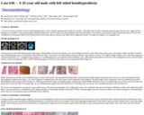
A 44 year-old female with familial Mediterranean fever (FMF), compound heterozygote with M680I and V726A mutations in the MEFV gene, on chronic colchicine therapy, cardiomyopathy with a low ejection fraction of 15-20%, and end stage renal disease on peritoneal dialysis was admitted for shortness of breath and syncopal episodes, complicated by pulseless electrical activity cardiac arrest. She otherwise did not have any acute or chronic neurologic symptoms (e.g. headaches, focal neurologic deficits, or seizures). The patient was resuscitated but developed respiratory failure, with suspected pneumonia and/or sepsis and was placed on antibiotics and inotropic agents; she developed a non ST elevation myocardial infarct. She subsequently had two additional episodes of pulseless electrical activity after which she was placed on extra-corporeal membrane oxygenation but without neurologic recovery. Computerized tomography (CT) showed no acute intracranial hemorrhage or mass effect. Given the poor prognosis, life-saving measures were withdrawn, and she expired.
(This case study was added to OER Commons as one of a …
(This case study was added to OER Commons as one of a batch of over 700. It has relevant information which may include medical imagery, lab results, and history where relevant. A link to the final diagnosis can be found at the end of the case study for review. The first paragraph of the case study -- typically, but not always the clinical presentation -- is provided below.)
The patient is a 44 year old female who was found to have an incidental heterogeneously enhancing splenic mass on CT scan. A follow up CT scan 2 years later demonstrated a solid appearing splenic mass with peripheral enhancement on the arterial phase and progressive enhancement on the portal venous phase. The size had increased from 1.6 cm to 2.9 cm so a splenectomy was performed.
(This case study was added to OER Commons as one of a …
(This case study was added to OER Commons as one of a batch of over 700. It has relevant information which may include medical imagery, lab results, and history where relevant. A link to the final diagnosis can be found at the end of the case study for review. The first paragraph of the case study -- typically, but not always the clinical presentation -- is provided below.)
A 44-year-old man with a past medical history of arterial hypertension, hypercholesterolemia, cigarette smoking (45 pack-years) and obesity (BMI 32.8) presented to our department with a 3-month history of right-sided facial numbness. Four weeks prior to admission he experienced a single episode of involuntary muscle movements on the left-side of his body. His neurologic exam was normal and initial laboratory results including CBC and blood chemistry were within normal range. A magnetic resonance imaging (MRI) scan of the patient's brain (Figure 1) showed a 7.3x4.9x3.6 cm, right fronto-parietal, extra-axial space-occupying lesion with lobulated contrast-enhancement and mild perifocal edema. The superior sagittal sinus was slightly compressed and the overlying cranium was infiltrated. The patient underwent angio-embolization of the lesion and two days later a right fronto-temporo-parietal craniectomy was performed. The tumor was resected subtotally, leaving a thin superficial infiltrative layer on eloquent cortex. The infiltrated cranium was reconstructed using polymethyl-methacrylate (PMMA) cranioplasty and the resected dura was replaced by a neuropatch. Postoperatively, an MRI of the spine and a lumbar puncture did not show any evidence for disease dissemination. The patient had no neurological deficit and underwent adjuvant radiation therapy of the tumor bed (36 Gy) and 2-years after diagnosis he was clinically and radiologically disease-free.
(This case study was added to OER Commons as one of a …
(This case study was added to OER Commons as one of a batch of over 700. It has relevant information which may include medical imagery, lab results, and history where relevant. A link to the final diagnosis can be found at the end of the case study for review. The first paragraph of the case study -- typically, but not always the clinical presentation -- is provided below.)
A 44-year-old man was hospitalized with an acute onset of giddiness, drunken gait and tendency to fall. He was apparently well until one week prior to presentation. He experienced these symptoms after drinking heavily at a party. There was no history of trauma, headache, vomiting or seizures.
(This case study was added to OER Commons as one of a …
(This case study was added to OER Commons as one of a batch of over 700. It has relevant information which may include medical imagery, lab results, and history where relevant. A link to the final diagnosis can be found at the end of the case study for review. The first paragraph of the case study -- typically, but not always the clinical presentation -- is provided below.)
The patient referred to the neurosurgeon in October 2015, complaining polyuria, polydipsia and polyphagia, associated with violent headache, unresponsive to anti-inflammatory therapy, vomit and loss of consciousness. After the onset of visual impairment in the left eye, the patient underwent MRI scan that revealed a giant irregular suprasellar mass (max diam 3.5 cm) moving from the level of the infundibular area within the third ventricle chamber; these lesion showed (Fig. 1A) strong and in-homogenous enhancement post-GAD and little calcified spots were noted within its bulk. Pituitary hormonal levels were within the normal range. In May 2016 the patient underwent surgery, by mean of endoscopic endonasal approach: at this time, due to the hard consistency, the tight adherences of the tumor and the narrow and deep corridor lesion has been only partially removed. As per protocol a second transcranial approach was scheduled, nevertheless three months later, hydrocephalus developed and urgent ventricle-peritoneal shunt procedure was required, although residual tumor was stable. Six months thereafter a new MRI disclosed a slight enlargement of tumor (volume increase was 30% more than the prior exam), so that transcranial transcortical-transventricular approach was adopted to remove the lesion. Extent of removal at that time was near-total (>90%), nevertheless, patient died few weeks later due to severe meningitis that complicated with multi-organ failure.
(This case study was added to OER Commons as one of a …
(This case study was added to OER Commons as one of a batch of over 700. It has relevant information which may include medical imagery, lab results, and history where relevant. A link to the final diagnosis can be found at the end of the case study for review. The first paragraph of the case study -- typically, but not always the clinical presentation -- is provided below.)
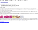
A 45-year-old woman presented to our institution with progressive upper and lower extremity proximal weakness of a few weeks' duration. 3 months prior, the patient received a diagnosis of systemic lupus erythematosus and started on hydroxychloroquine; she had been treated with low dose prednisone for her weakness for the last 3 weeks. Physical examination showed severe proximal weakness, grade 2/5, in the bilateral upper and lower extremities with retained strength in all other muscle groups. The patient had an erythematous rash on her face, chest, and upper back. There was no clinical evidence of involvement of other organs.
(This case study was added to OER Commons as one of a …
(This case study was added to OER Commons as one of a batch of over 700. It has relevant information which may include medical imagery, lab results, and history where relevant. A link to the final diagnosis can be found at the end of the case study for review. The first paragraph of the case study -- typically, but not always the clinical presentation -- is provided below.)
A 45-year-old man presented with left-sided hemihypesthesia, which remitted spontaneously within two months. 18 months later the same symptoms appeared again but were now aggravated by hemiparesis, dysarthria, ataxia and neurogenic bladder dysfunction, which finally led to pyelonephritis and acute renal failure. The patient's past medical, surgical and family history were all non-contributory. Cerebrospinal fluid (CSF) showed normal cell counts, glucose, protein, IgG index and no oligoclonal bands. Cytology was negative for malignant cells.
(This case study was added to OER Commons as one of a …
(This case study was added to OER Commons as one of a batch of over 700. It has relevant information which may include medical imagery, lab results, and history where relevant. A link to the final diagnosis can be found at the end of the case study for review. The first paragraph of the case study -- typically, but not always the clinical presentation -- is provided below.)
In 1999, at the age of 41 this man developed focal seizures in his right arm. The neurological examination was otherwise normal. He was in good health and his medical history was devoid of underlying disease. Cranial MRI revealed a homogeneously contrast enhancing lesion with microcalcifications in the left frontal lobe (Figs. 1 and 2). One year later the patient decided to have stereotactic biopsy. Surgically induced artifacts and small sample size aggravated tumor classification at that time. Differential diagnoses included oligodendroglioma WHO II and diffuse astrocytoma WHO II. In January 2003, at the age of 45, the patients developed weakness in his right arm and seizure frequency increased despite medication. Tumor size had considerably increased. Surgery was offered and the patient decided to have the lesion removed through a left frontal craniotomy. The postoperative course, was unremarkable, the weakness of the right arm disappeared.
(This case study was added to OER Commons as one of a …
(This case study was added to OER Commons as one of a batch of over 700. It has relevant information which may include medical imagery, lab results, and history where relevant. A link to the final diagnosis can be found at the end of the case study for review. The first paragraph of the case study -- typically, but not always the clinical presentation -- is provided below.)
This 45 year old man presented to the emergency room with a history of a small quarter - sized midline tongue lesion present since childhood, which had recently increased in size to that of a golf ball and was associated with spontaneous drainage of a dark brown fluid. There was no mucosal ulceration, or history of dyspnea, odynophagia or bleeding.
(This case study was added to OER Commons as one of a …
(This case study was added to OER Commons as one of a batch of over 700. It has relevant information which may include medical imagery, lab results, and history where relevant. A link to the final diagnosis can be found at the end of the case study for review. The first paragraph of the case study -- typically, but not always the clinical presentation -- is provided below.)
A 45-year-old man presented to us with headache and vomiting for15 years ago. He was diagnosed as having obstructive hydrocephalus due to a pineal region tumour. A ventriculoperitoneal shunt was inserted to relieve hydrocephalus with symptomatic relief. He was followed up with regular MRI scans. Serial imaging showed slow progression in tumor size when he was 60 years old. Neurological examination revealed Parinaud sign with upward gaze palsy. The MRI with gadolinium showed an mildly enhancing lobulated tumor in the region of the superior tectum measuring 22.5mm x 21mm x 20mm (Figs. 1 and 2). The lesion at the superior tectum and the pineal region showed heterogeneous T1-hyperintense signal on precontrast images (Fig. 1). It exhibited very slow growth, with a gradual increase in size over 15 years. There was mild mass effect to the cerebral aqueduct with indentation of the pineal gland (Fig. 3). However, there was no hydrocephalus at that time. In view of symptomatic progression of tumor size, he underwent a near total tumour excision via infratentorial supracerebellar approach.
(This case study was added to OER Commons as one of a …
(This case study was added to OER Commons as one of a batch of over 700. It has relevant information which may include medical imagery, lab results, and history where relevant. A link to the final diagnosis can be found at the end of the case study for review. The first paragraph of the case study -- typically, but not always the clinical presentation -- is provided below.)
A 46-year-old woman was admitted to our hospital with chief complaint of numbness of right lower limb lasting 1 year as well as left leg ache lasting 5 months. The symptoms were gradually developing. There was no history of trauma, drug use or any physical exertion. Neurological examination confirmed sense of pain and temperature subsided in left upper limb, left side of body and right lower limb. Radiating pain happened at the left lap and rear waist, and the tendon reflex weakened slightly at left upper limb and right lower limb. A spinal MRI revealed a heterogeneous intensity enhancing 3.0×1.0×1.0cm mass involving T5 through T7 (Figure 1a, b); it was hypointense on T1 (Figure 1c) and hyperintense on T2-weighted sequences (Figure 1d). Short time inversion recovery (STIR) image showed minimal perilesional edema (Figure 1e). The patient subsequently underwent a near total tumour excision via T5-T7 laminectomy approach. Then she underwent craniospinal irradiation and temozolomide chemotherapy. Postoperative follow-up was uneventful with good control in 12 months.
No restrictions on your remixing, redistributing, or making derivative works. Give credit to the author, as required.
Your remixing, redistributing, or making derivatives works comes with some restrictions, including how it is shared.
Your redistributing comes with some restrictions. Do not remix or make derivative works.
Most restrictive license type. Prohibits most uses, sharing, and any changes.
Copyrighted materials, available under Fair Use and the TEACH Act for US-based educators, or other custom arrangements. Go to the resource provider to see their individual restrictions.