(This case study was added to OER Commons as one of a …
(This case study was added to OER Commons as one of a batch of over 700. It has relevant information which may include medical imagery, lab results, and history where relevant. A link to the final diagnosis can be found at the end of the case study for review. The first paragraph of the case study -- typically, but not always the clinical presentation -- is provided below.)
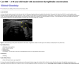
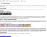
The patient is a 46 year old female being evaluated for bariatric surgery. During an endoscopy procedure, a raised, "yellowish" nodule (size not specified) was identified at the mid esophagus, 30 cm from the incisors (image 1). An endoscopic mucosal resection (EMR) was performed. Grossly the lesion was described as a 0.6 x 0.4 cm yellow-red, ulcerated and centrally loculated lesion (image 2). H&E images of the EMR specimen are provided in images 3, 4, 5 and 6. PASD staining is shown in images 7 and 8. Immunohistochemistry is shown for S100 is in image 9 and inhibin in image 10.
(This case study was added to OER Commons as one of a …
(This case study was added to OER Commons as one of a batch of over 700. It has relevant information which may include medical imagery, lab results, and history where relevant. A link to the final diagnosis can be found at the end of the case study for review. The first paragraph of the case study -- typically, but not always the clinical presentation -- is provided below.)
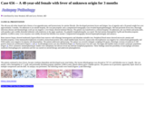
A 46 year old white female presented to the endocrine clinic for followup on hypothyroidism and a palpable thyroid nodule. Ultrasound of her thyroid on 09/02/03, revealed multiple nodules including a nodule in the upper right pole measuring 7 x 9mm, a nodule in the left aspect of the isthmus measuring 9 x 4mm (Figure 1), and a nodule in the mid pole of the right thyroid measuring 5 x 5mm. An ultrasound guided fine needle aspiration was performed on the nodule near the isthmus which showed papillary carcinoma. A total thyroidectomy with central lymph node dissection was performed on 09/26/XX which showed papillary carcinoma with one positive lymph node, T1N1MX.
(This case study was added to OER Commons as one of a …
(This case study was added to OER Commons as one of a batch of over 700. It has relevant information which may include medical imagery, lab results, and history where relevant. A link to the final diagnosis can be found at the end of the case study for review. The first paragraph of the case study -- typically, but not always the clinical presentation -- is provided below.)
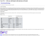
Patient is a 46-year-old female with no significant past medical history. She presented to an outside hospital with a three day history of shortness of breath and palpitations. A chest x-ray revealed bilateral lung infiltrates. She was subsequently intubated for respiratory failure and sedated. She also had black, tarry stools and bloody orogastric tube output, and was subsequently admitted to an outside hospital intensive care unit for sepsis. Reported initial complete blood count values included a hemoglobin value of 7.4 gm/dl, platelet count of 13x10E+9/L, and white blood cell count of 10.3x10E+9/L, The peripheral blood differential reportedly demonstrated 15% blasts. She was transferred to the intensive care unit at UPMC for further management. A bone marrow biopsy was performed for further evaluation.
(This case study was added to OER Commons as one of a …
(This case study was added to OER Commons as one of a batch of over 700. It has relevant information which may include medical imagery, lab results, and history where relevant. A link to the final diagnosis can be found at the end of the case study for review. The first paragraph of the case study -- typically, but not always the clinical presentation -- is provided below.)
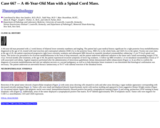
A 46-year-old man presented with a 2 week history of bilateral lower extremity numbness and tingling. The patient had a past medical history significant for a right posterior fossa medulloblastoma diagnosed at the age of 24, treated with total resection and craniospinal radiation (5040 cGy to the posterior fossa, 3960 cGy to the whole brain, and 3420 cGy to the spine). Twenty-one years later, at the age of 45, the patient experienced progressive right lower extremity weakness and subsequent MRI showed an expansile intradural extramedullary enhancing 1.3 cm T5 level spinal cord lesion. This lesion was presumed to be recurrent medulloblastoma in the form of drop metastasis, and the patient underwent additional radiation to the tumor and a small surrounding margin (3750 cGy). Several months after treatment, at the age of 46, the patient experienced recurrent symptoms of lower leg weakness. A follow-up MRI revealed a 1.4 cm intradural extramedullary lesion at T7 with associated cord edema. Sagittal sequences performed after the administration of intravenous gadolinium chelate demonstrated subtle enhancement (Figure 1). In an effort to confirm the diagnosis of recurrent meduloblastoma and rule out radiation necrosis or a second malignancy as well as to help determine future treatment it was determined that histological confirmation was necessary. The patient underwent an uneventful thoracic laminectomy at T6-T7 with subtotal resection of the intradural lesion.
(This case study was added to OER Commons as one of a …
(This case study was added to OER Commons as one of a batch of over 700. It has relevant information which may include medical imagery, lab results, and history where relevant. A link to the final diagnosis can be found at the end of the case study for review. The first paragraph of the case study -- typically, but not always the clinical presentation -- is provided below.)
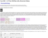
A 39-year-old man was admitted in 2009 following epileptic seizure associated with visual impairment. MRI revealed an expansive and infiltrative lesion in the right parieto-occipital lobe with a cortico-subcortical localization. This lesion was hypointense in T1-weighted images and showed no contrast enhancement after gadolinium injection. Methionine-positron emission tomography (MET-PET) revealed high methionine uptake in this lesion. Partial surgical resection of the lesion was performed. No adjuvant therapy was administered at this moment. MRI images showed progression of the lesion in 2014 but the patient refused surgical intervention and other therapies. In 2017, following painful headache associated with neurological deficit, the MRI suggested progressive neoplasm in the right parieto-occipital lobe (Figure 1) with areas of enhancement on post contrast MRI. MET-PET revealed heterogeneous increased metabolic uptake, suggesting progression of the disease. New surgery was performed.
(This case study was added to OER Commons as one of a …
(This case study was added to OER Commons as one of a batch of over 700. It has relevant information which may include medical imagery, lab results, and history where relevant. A link to the final diagnosis can be found at the end of the case study for review. The first paragraph of the case study -- typically, but not always the clinical presentation -- is provided below.)
Patient is a 47 year old female who presented to an outpatient clinic with the chief compliant of thigh swelling due to what was thought to be a blood clot. She received a MRI scan which showed an incidental mass in the contralateral thigh. A more thorough MRI confirmed the 7cm heterogenous lesion in her left vastus intermedius. At this point of time, it was felt to be a sarcoma and a biopsy was performed.
(This case study was added to OER Commons as one of a …
(This case study was added to OER Commons as one of a batch of over 700. It has relevant information which may include medical imagery, lab results, and history where relevant. A link to the final diagnosis can be found at the end of the case study for review. The first paragraph of the case study -- typically, but not always the clinical presentation -- is provided below.)
A 47-year-old male patient presented with a fluctuating hearing impairment in his left ear over the past 5 years. Tinnitus or vertigo was not observed. Audiometric analysis showed an inner ear deficit of 50 dB between 1500 and 6000 kHz on the left side. Hearing in the right ear was normal. The facial nerve was clinically and by means of electrophysiological testing without pathological findings. BEAP (brainstem evoked auditory potentials) revealed a latency increase between J1 and J3 up to 2.5 ms for the left side, whereas only 2.3 ms on the right side. MRI (magnetic resonance imaging) scanning showed a tumor of the cerebellopontine angle in the left inner auditory canal (IAC) of 1.2 x 0.7 x 0.9 cm in size (Figure 1). After application of contrast media, the tumor showed clear signal enhancement. The tumor was entirely removed by a transtemporal approach to the IAC. Surgical exploration found the cochlear nerve embedded in a tumorous mass, whereas the vestibular and the facial nerve were normal (Figure 2, vestibular nerve arrow A and cochlear nerve with tumor arrow B). Nerve and tumor (Figure 3) were removed and sent to histopathological examination. The patient lost his hearing after the operation due to the removal of the cochlear nerve, whereas a regular postoperative vestibular function was observed. The postoperative course as well as the 5-year follow-up examination was unremarkable and control MRI scanning showed no recurrence.
(This case study was added to OER Commons as one of a …
(This case study was added to OER Commons as one of a batch of over 700. It has relevant information which may include medical imagery, lab results, and history where relevant. A link to the final diagnosis can be found at the end of the case study for review. The first paragraph of the case study -- typically, but not always the clinical presentation -- is provided below.)
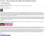
A 47 year-old man presented with left extremity weakness for 1 year, which had worsened significantly over the past 40 days. 23 years ago he suffered head trauma with loss of consciousness for 30 minutes and was diagnosed with a concussion. Two weeks later he had his first generalized seizure, and continued to have about one seizure per week. His seizures were not well-controlled on phenytoin. Fifteen years ago, the patient underwent brain CT scan, revealing right frontal encephalomalacia (Figure 3). Epilepsy surgery was performed and silver clips were deployed for hemostasis. Following the surgery and change of medications to carbamazepine he continued to suffer seizures about one or twice a month. At this current presentation for left extremity weakness, the neurological examination discovered decreased muscle strength of the left side. CT scan revealed a huge hypodense lesion in the right frontal lobe with remarkable mass effect (Figure 1), but without distinct contrast-enhancement (Figure 2). Hyperdense foci could be seen within the mass, which were ascertained to be the silver clips used in his surgery. MRI scan could not be performed due to these clips. A right frontal craniotomy was performed and a large tumor was removed and submitted for pathology.
(This case study was added to OER Commons as one of a …
(This case study was added to OER Commons as one of a batch of over 700. It has relevant information which may include medical imagery, lab results, and history where relevant. A link to the final diagnosis can be found at the end of the case study for review. The first paragraph of the case study -- typically, but not always the clinical presentation -- is provided below.)
A 47-year-old white, right-handed man presented with abrupt onset of left leg numbness while he was at work. He walked around to try to restore circulation when it suddenly became paralyzed and noticed his left arm and leg began to contract rhythmically and involuntarily. There was no report of eye deviation, face involvement, or loss of consciousness. At the time of arrival to the hospital, the movements had stopped. He had no prior history of seizure or stroke. His past medical history was significant for migraine headaches, sinus allergies, chronic uveitis and a history of parathyroidectomy in the past. He had an episode of severe uveitis at age eight with bilateral eye redness and soreness and was treated with steroids and Cytoxan. His medications were oxymetazoline nasal spray for nasal congestion and daily prednisolone eyedrops. He has a 20-year history of tobacco use and drinks alcohol socially. He denied any unusual exposure history and to his knowledge had never been exposed to tuberculosis. His family history was notable for a mother with breast cancer, and sister with Sjögren's syndrome who had uveitis as well.
(This case study was added to OER Commons as one of a …
(This case study was added to OER Commons as one of a batch of over 700. It has relevant information which may include medical imagery, lab results, and history where relevant. A link to the final diagnosis can be found at the end of the case study for review. The first paragraph of the case study -- typically, but not always the clinical presentation -- is provided below.)
A 47-year-old woman was admitted to the hospital with a 4-month history of frontal headache that had radiated to her neck. Physical and neurological examinations did not demonstrate any abnormalities.
(This case study was added to OER Commons as one of a …
(This case study was added to OER Commons as one of a batch of over 700. It has relevant information which may include medical imagery, lab results, and history where relevant. A link to the final diagnosis can be found at the end of the case study for review. The first paragraph of the case study -- typically, but not always the clinical presentation -- is provided below.)
This 48-year-old white female had a history of an appendectomy and hysterectomy for uterine fibroids. She developed persistent fevers and fatigue, loss of appetite and a 20-pound weight loss over approximately 3 months. On admission to an outside hospital, she was pancytopenic and a computerized tomography scan revealed hepatosplenomegaly. She had persistent fevers but a thorough infectious disease and rheumatologic work up was negative. Liver biopsy showed minimal fibrosis. The patient was transferred to a referral hospital. On admission, she was febrile and tachycardic, with jaundice and a mildly distended abdomen with tenderness in the upper quadrants. No palpable lymphadenopathy was noted. She had anemia (hemoglobin 9 g/dl) and thrombocytopenia (platelets 32,000/cu mm). Liver enzymes were mildly elevated but bilirubin was normal. Computerized tomography showed hepatosplenomegaly and anasarca.
(This case study was added to OER Commons as one of a …
(This case study was added to OER Commons as one of a batch of over 700. It has relevant information which may include medical imagery, lab results, and history where relevant. A link to the final diagnosis can be found at the end of the case study for review. The first paragraph of the case study -- typically, but not always the clinical presentation -- is provided below.)
The patient is a 48 year old Caucasian male with a past medical history of hypertension, gastroesophageal reflux disease, irritable bowel syndrome, chronic headaches, anxiety, depression, chronic low back pain secondary to spinal stenosis status post L3 through L5 laminectomies in 2009, and end-stage osteoarthritis in bilateral hips status post total left hip (March) and total right hip (September) arthroplasties with metal-on-metal prostheses in 2008. The patient worked in the construction industry for many years and performed daily tasks that resulted in his chronic hip disease, including heavy lifting and prolonged kneeling. Following the left hip replacement, his pain was vastly improved and he did well with no complications. However, following the right hip replacement, he noted increased bilateral hip pain, right more than left. In April 2012, he presented to an outpatient clinic with these complaints, in addition to complaints of increasing confusion, headaches, blurry vision for about two weeks, and short-term memory loss. Radiologic imaging was performed at that time, but failed to reveal signs of joint failure. Interestingly, a blood cobalt level was drawn and returned 11.4 µg/L (reference range: <1.8 µg/L). He subsequently underwent a revision of the right hip in October 2012, replacing the metal-on-metal joint with a non-metal surface prosthesis. A repeat blood cobalt level was drawn after the surgery and returned 5 µg/L. He remained clinically stable following the right hip revision, but in February 2013 he presented to an outside hospital after he sustained a traumatic fall in his basement. He attributed the fall to symptoms of progressive cognitive decline, including confusion, lethargy, short-term memory loss, and depression, which he noted had worsened since his hip procedures in 2008. Computed tomography (CT) and magnetic resonance imaging (MRI) scans of the head did not reveal significant acute pathology, an electroencephalogram (EEG) was also negative for seizure-like activity or any other pathologic activity in the brain, and all other work-ups were essentially negative. On admission to the hospital, he stated that he had a history of elevated blood cobalt levels, but the patient's previous laboratory studies were not available for review by his clinicians. A repeat blood cobalt level was drawn during his hospitalization and returned 3.1 µg/L. The patient was eventually discharged from the hospital with follow-up at an outpatient toxicology clinic for further evaluation.
(This case study was added to OER Commons as one of a …
(This case study was added to OER Commons as one of a batch of over 700. It has relevant information which may include medical imagery, lab results, and history where relevant. A link to the final diagnosis can be found at the end of the case study for review. The first paragraph of the case study -- typically, but not always the clinical presentation -- is provided below.)
This 48 year old male presented in October 2000 with sudden (<12 hours) right sided weakness. His admission blood pressure was 178/107. Nervous system examination found reduced visual acuity but no double vision or visual field defects. Right sided power was reduced to 0/5 (right arm) and 3/5 (right leg), with normal tone, reduced bulk and increased reflexes. Plantars were bilaterally up-going with ankle clonus. Admission CT scan showed a 3cm (transverse) x 2cm (sagittal) deep left intracerebral hematoma with surrounding edema. The occipital horns of the lateral ventricles were compressed by bilateral hypointense white matter lesions (figure 1). The patient became unresponsive the day after admission, with bilateral fixed pupils and a repeat CT showed hematoma expansion with new onset left uncal herniation. The bilateral white matter hypointensities were unchanged. Despite further treatment, on this day, with family consent, the patient was extubated and expired. Consent was given to perform a complete autopsy.
(This case study was added to OER Commons as one of a …
(This case study was added to OER Commons as one of a batch of over 700. It has relevant information which may include medical imagery, lab results, and history where relevant. A link to the final diagnosis can be found at the end of the case study for review. The first paragraph of the case study -- typically, but not always the clinical presentation -- is provided below.)
A 48- year- old female presented with right sided moderate to severe headache for 2 years. She also had difficulty in right lateral vision for 4-5 months. For the last 2 months she developed difficulty in swallowing liquids and solids. The patient was unable to walk properly and had a tendency to fall on her right side for last 15 days. Her past history was significant for hypertension for which she was taking antihypertensives. On neurological examination, the patient was conscious and oriented. There was right sided cranial nerve palsy which includes III, IV, V, IX, X and XI nerves. The cerebellar signs were positive on right side. The MRI showed a mass lesion probably in relation to trigeminal nerve on right side showing extension anteriorly to Meckels cave and posteriorly to the cerebellopontine angle. The lesion was hyperintense on T2 and hypointense on T1 weighted images. After the administration of intravenous contrast gadolinium DTPA, the lesion showed moderate to intense homogenous enhancement. The lesion extended into the cavernous sinus and petrous bone on the right side. It measured 5.7x3.1x5.5 cm. The lesion was causing marked indentation of the pons and pontocerebellar junction on the right side. The rest of the brain parenchyma revealed no abnormal signal intensity. These findings were suggestive of a right trigeminal nerve schwannoma showing extension and mass effect or a petrous meningioma. Patient underwent suboccipital retrosigmoid craniotomy with excision of the mass lesion.
(This case study was added to OER Commons as one of a …
(This case study was added to OER Commons as one of a batch of over 700. It has relevant information which may include medical imagery, lab results, and history where relevant. A link to the final diagnosis can be found at the end of the case study for review. The first paragraph of the case study -- typically, but not always the clinical presentation -- is provided below.)
The patient was a 48-year-old woman with a one-year history of back pain, who recently developed right upper quadrant pain that radiated to her side and back. The past medical history included cervical cancer in 1983, bipolar disorder, fibromyalgia, and Hashimoto's thyroiditis. Medications at presentation listed Synthroid, lithium, and Aviane (ethinyl estradiol and levonorgestrel). She had been on birth control pills for the past 20 years.
(This case study was added to OER Commons as one of a …
(This case study was added to OER Commons as one of a batch of over 700. It has relevant information which may include medical imagery, lab results, and history where relevant. A link to the final diagnosis can be found at the end of the case study for review. The first paragraph of the case study -- typically, but not always the clinical presentation -- is provided below.)
A 48-year-old right handed female presented with a 6 month history of progressive decrease of sensation in right upper and lower extremities following a fall after returning from a golf course. She attributed that fall to the loss of sensation, which improved spontaneously over 10 minutes with some residual numbness in the right leg. Her symptoms progressed and she developed difficulty in walking and lifting her right leg. She also complained of neck discomfort and chronic headaches. Neurological examination revealed mild patchy loss of pinprick and light touch sensation in the right upper and lower extremity and trunk with no consistent dermatomal distribution. There was diminished vibration sensation in the right knee. Deep tendon reflex was absent in the right ankle. Plantar reflexes were downgoing. Her gait was stiff with mild circumduction of the right leg. Rest of the motor and cranial nerve examinations were grossly normal.
(This case study was added to OER Commons as one of a …
(This case study was added to OER Commons as one of a batch of over 700. It has relevant information which may include medical imagery, lab results, and history where relevant. A link to the final diagnosis can be found at the end of the case study for review. The first paragraph of the case study -- typically, but not always the clinical presentation -- is provided below.)
A 49-year old female with no significant past medical history except for psoriasis presented to our department complaining of increasing neck pain over 4 weeks accompanied by hoarseness and dysphagia. In addition, she reported general fatigue as well as problems with fine motor skills of her right arm and right leg. On MRI we found a left anterolateral extra-axial space-occupying lesion (45 x 32 x 32 mm) of the foramen magnum that shifted the brain stem to the right, another small para-torcular (8mm) extra-axial lesion over the left cerebellar convexity and an extra-axial mass over the right sylvian fissure (shown in Figs. 1a and 1b). Considering the clinical symptoms of the patients, we opted for surgery of the foramen magnum tumor and the small peritorcular lesion of the left cerebellar convexity due to its proximity. After angioembolization of the supratentorial tumor and foramen magnum mass, a far lateral suboccipital craniotomy was performed on the left side in park bench position. Surgery was complicated by the difficult anatomy with the lower cranial nerves and brain stem perforators of the vertebral artery running on the surface of the tumor. Using a combination of intratumoral ultrasonic aspiration and peritumoral microdissection, the foramen magnum mass could be removed completely and the dural attachment was coagulated (corresponding to a Simpson grade II resection in meningioma surgery). The small para-torcular lesion had a clear arachnoidal plane and could be completely resected without complications. The patient had no new postoperative neurological deficits and the postoperative MRI showed a complete resection of both lesions ( Figs. 1a and 1b). At three months follow-up, the preoperative symptoms recovered completely. The foramen magnum mass was diagnosed as a WHO grad I meningothelial meningioma - the diagnosis of the smaller cerebellar para-torcular mass is discussed below in detail.
(This case study was added to OER Commons as one of a …
(This case study was added to OER Commons as one of a batch of over 700. It has relevant information which may include medical imagery, lab results, and history where relevant. A link to the final diagnosis can be found at the end of the case study for review. The first paragraph of the case study -- typically, but not always the clinical presentation -- is provided below.)
A 48 years-old patient without significant medical history or without bleeding disorder is addressed to our institution for acute headache and bitemporal hemianopsia. No hormonal secretion was detected, and the patient had a pituitary insufficiency with loss of libido, diffuse hair loss, asthenia and slowing. An MRI showed a 47mm pituitary lesion occupying the sella turcica with central necrosis. This lesion was hypointense in T1 sequence, enhanced with gadolinium injection and hyperintense in T2 sequence (Fig.1). Then, a transsphenoidal excision surgery was decided. A first surgery was performed, but the resection of the tumor lead to an extensive bleeding and the tumor could not be resected entirely. Three days later, because of incomplete resection a second excision procedure was decided leading to an extensive bleeding and a subtotal surgical resection. The visual symptoms of the patient improved quickly after the surgery.
(This case study was added to OER Commons as one of a …
(This case study was added to OER Commons as one of a batch of over 700. It has relevant information which may include medical imagery, lab results, and history where relevant. A link to the final diagnosis can be found at the end of the case study for review. The first paragraph of the case study -- typically, but not always the clinical presentation -- is provided below.)
A 49-year-old man presented with a two-month history of headache and recent twenty pound weight loss. He was diagnosed with cluster headaches at an outside hospital and treated with triptans. One week after the initial onset of headache, he developed right jaw and facial pain with facial weakness, accompanied by inability to fully close his right eye and mouth. He was given one dosage of steroids at an outside hospital for cluster headache with full resolution of his symptoms. However, a week later, his right facial weakness recurred and persisted. About four weeks later, he complained about similar jaw/facial pain and weakness of his left side, which appeared to be more severe than those of his right side. He also developed occasional diplopia on far gaze and left lateral gaze. He did not experience any changes in hearing or taste. Magnetic resonance imaging of brain revealed abnormal enhancement of the third, fifth, seventh and eighth cranial nerves with various degree of diffuse enlargement (arrows pointing to trigeminal nerves in Figure 1). Patchy enhancement of the nerve roots in the distal lumbar region was also noted. A gallium whole body scan did not reveal any significant abnormality. Repeated CSF studies showed low glucose levels, elevated protein levels and elevated cell counts. Extensive peripheral blood and CSF work-up for infectious disease, autoimmune, demyelinating disorders and neoplasia were negative except for a positive T-cell lymphotropic virus type 1 (HTLV-1) antibody. His HIV-1/2 ELISA was negative. Peripheral blood flow cytometry showed mature WBCs with slightly elevated CD4:CD8 ratio. CSF flow cytometry revealed large granular T-cell lymphocytosis consisting of CD3/CD56/CD57+ lymphocytes (>97% of gated lymphocytes), which was interpreted as due to a reactive process. A working diagnosis of neurosarcoidosis was made and the patient was treated with prednisone 60 mg daily for two weeks followed by dexamethasone 4 mg daily for another two weeks. However, the patient's symptoms worsened, leading to heightened suspicion of a malignant neoplasm. Dexamethasone was discontinued and he subsequently underwent biopsy of the enlarged left infraorbital nerve (Figure 2, arrows).
(This case study was added to OER Commons as one of a …
(This case study was added to OER Commons as one of a batch of over 700. It has relevant information which may include medical imagery, lab results, and history where relevant. A link to the final diagnosis can be found at the end of the case study for review. The first paragraph of the case study -- typically, but not always the clinical presentation -- is provided below.)
A 49-year-old female, who suffered trauma to the left fronto-parietal region from a gymnasium weight two years previously, presented with a 12-month history of an enlarging lump in the same area. There were no associated visual, sensory or neurological symptoms. On examination there was a large smooth, non-tender, bony hard mass measuring 8 x 6 centimetres at its base, with mild to moderate overlying alopecia. It was non-pulsatile with no bruits. The lesion was percussion dull and there was no regional lymphadenopathy.
No restrictions on your remixing, redistributing, or making derivative works. Give credit to the author, as required.
Your remixing, redistributing, or making derivatives works comes with some restrictions, including how it is shared.
Your redistributing comes with some restrictions. Do not remix or make derivative works.
Most restrictive license type. Prohibits most uses, sharing, and any changes.
Copyrighted materials, available under Fair Use and the TEACH Act for US-based educators, or other custom arrangements. Go to the resource provider to see their individual restrictions.