(This case study was added to OER Commons as one of a …
(This case study was added to OER Commons as one of a batch of over 700. It has relevant information which may include medical imagery, lab results, and history where relevant. A link to the final diagnosis can be found at the end of the case study for review. The first paragraph of the case study -- typically, but not always the clinical presentation -- is provided below.)
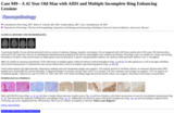
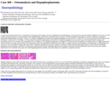
A previously healthy 42-year-old man presented with two weeks of confusion, lethargy, headache, and malaise. He was diagnosed with AIDS three months prior (CD4 count 198 cells/microliter; viral load 87,100 copies/mL) and was on trimethoprim/sulfamethoxazole prophylaxis but had not started highly active antiretroviral therapy. Neurologic exam was notable for a sleepy man lacking orientation to location or date, perseveration on simple phrases and overall paucity of speech, and generalized weakness. He could not stand unassisted. Reflexes were 3+ throughout.
(This case study was added to OER Commons as one of a …
(This case study was added to OER Commons as one of a batch of over 700. It has relevant information which may include medical imagery, lab results, and history where relevant. A link to the final diagnosis can be found at the end of the case study for review. The first paragraph of the case study -- typically, but not always the clinical presentation -- is provided below.)
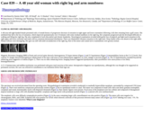
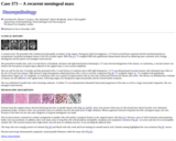
A 48-year-old right handed female presented with a 6 month history of progressive decrease of sensation in right upper and lower extremities following a fall after returning from a golf course. She attributed that fall to the loss of sensation, which improved spontaneously over 10 minutes with some residual numbness in the right leg. Her symptoms progressed and she developed difficulty in walking and lifting her right leg. She also complained of neck discomfort and chronic headaches. Neurological examination revealed mild patchy loss of pinprick and light touch sensation in the right upper and lower extremity and trunk with no consistent dermatomal distribution. There was diminished vibration sensation in the right knee. Deep tendon reflex was absent in the right ankle. Plantar reflexes were downgoing. Her gait was stiff with mild circumduction of the right leg. Rest of the motor and cranial nerve examinations were grossly normal.
(This case study was added to OER Commons as one of a …
(This case study was added to OER Commons as one of a batch of over 700. It has relevant information which may include medical imagery, lab results, and history where relevant. A link to the final diagnosis can be found at the end of the case study for review. The first paragraph of the case study -- typically, but not always the clinical presentation -- is provided below.)
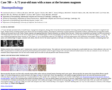
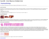
A 53-year-old white male with a history of alcohol abuse and pancreatitis presented to the emergency department with a 6-month history of nausea, vomiting and weight loss. One month prior, he had been diagnosed with cirrhosis of the liver, and subsequently hospitalized twice with removal of two and then five liters of ascites fluid. He developed significant abdominal pain associated with nausea and vomiting. He also had difficulty tolerating oral intake and constipation. He was not eating but sustaining himself on fluids. On physical examination, the patient was not jaundiced, but his abdomen was distended with a positive fluid wave. A firm mass was noted in the left mid epigastrium judged consistent with splenomegaly. His total bilirubin was 0.5 (direct 0.1), alkaline phosphatase 102, ALT 27, AST 17, albumin 3.4, total protein 6.3, prothrombin time 9.4, INR 1, partial thromboplastin time 33.8, amylase 257 and lipase 1130. He was admitted with a diagnosis of acute pancreatitis. He was made n.p.o. and given IV fluids.
(This case study was added to OER Commons as one of a …
(This case study was added to OER Commons as one of a batch of over 700. It has relevant information which may include medical imagery, lab results, and history where relevant. A link to the final diagnosis can be found at the end of the case study for review. The first paragraph of the case study -- typically, but not always the clinical presentation -- is provided below.)
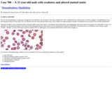
This 55 year-old gentleman is status-post laryngectomy for squamous cell carcinoma of the larynx diagnosed in 2010, complicated by a tracheostomy occlusion, leading to cardiopulmonary arrest and subsequent hypoxic brain injury. He was admitted with new-onset altered mental status, after weakness for approximately 6 months. On admission he was found to be severely malnourished.
(This case study was added to OER Commons as one of a …
(This case study was added to OER Commons as one of a batch of over 700. It has relevant information which may include medical imagery, lab results, and history where relevant. A link to the final diagnosis can be found at the end of the case study for review. The first paragraph of the case study -- typically, but not always the clinical presentation -- is provided below.)
The patient was a 58-year-old African American woman with severe static encephalopathy and cerebral palsy both presumptively related to prenatal/perinatal brain injury. Additional neurological diagnoses included an ill-defined seizure history, bipolar affective disorder, and medication-related tardive dyskinesia. The patient's other co-morbidities were non-contributory. She was a permanent resident of a long-term care facility, where she expired unexpectedly. An autopsy was requested.
(This case study was added to OER Commons as one of a …
(This case study was added to OER Commons as one of a batch of over 700. It has relevant information which may include medical imagery, lab results, and history where relevant. A link to the final diagnosis can be found at the end of the case study for review. The first paragraph of the case study -- typically, but not always the clinical presentation -- is provided below.)
A 73 year-old male presented with a 6 month history of progressive tetraparesis, during which he had deteriorated markedly from being mobile and performing in a steel band to completely bed-bound. Medical history included ischemic heart disease and type II diabetes mellitus. Neurological examination revealed symmetrical pyramidal weakness, pathologically increased deep tendon reflexes and increased tone in all 4 limbs. Plantar response was upgoing bilaterally. Pinprick-sensation was reduced below the neck. Cranial nerve examination was normal. An MRI scan showed a well-demarcated extra-axial mass on the dorsal surface of the lower medulla/upper cervical cord extending from the level of the clivus to the body of the axis inferiorly (figures 1, 2, 3 and 4). The mass exhibited heterogeneous hyper-intensity on T2W (figure 1), low-to-intermediate signal on T1W (figure 2), and florid enhancement with Gadolinium contrast-agent (figures 3 and 4). The spinal cord exhibited high T2-signal, consistent with edema, from the level of the tumour superiorly to the body of C5 inferiorly. There was no bony erosion or sclerosis, or enhancing dural 'tail'.
(This case study was added to OER Commons as one of a …
(This case study was added to OER Commons as one of a batch of over 700. It has relevant information which may include medical imagery, lab results, and history where relevant. A link to the final diagnosis can be found at the end of the case study for review. The first paragraph of the case study -- typically, but not always the clinical presentation -- is provided below.)
This 77-year-old white male had insulin-dependent diabetes mellitus, hyperlipidemia, peripheral vascular disease, hypothyroidism, peptic ulcer disease attributed to non-steroidal anti-inflammatory drug use, and a remote smoking history.
(This case study was added to OER Commons as one of a …
(This case study was added to OER Commons as one of a batch of over 700. It has relevant information which may include medical imagery, lab results, and history where relevant. A link to the final diagnosis can be found at the end of the case study for review. The first paragraph of the case study -- typically, but not always the clinical presentation -- is provided below.)
A 7-year-old Chinese boy with a history of bronchial asthma had upper respiratory tract infection for the preceding two weeks treated by a pediatrician. He then presented with confusion and incoherent speech on the day of admission. He was afebrile, with the Glasgow coma scale (GCS) of 14/15 (E4V4M6) and normal physical findings. Investigations showed normal complete blood counts, blood glucose, renal and liver function tests except for mildly elevated ALT at 34 IU/L (5-25 IU/L). His blood ammonia, lactate and pyruvate levels were normal. The CT brain showed no space occupying lesion or cerebral edema. Lumbar puncture showed an opening pressure of 12cm water, CSF glucose 3.1mmol/L, protein 1.25 gm/L (0.15-0.45 gm/L), RBC 2/mm3 and WBC 2/mm3. The gram stain showed no organisms. The nasopharyngeal aspirate for rapid antigen test for both influenza A and B were negative. About 12 hr after admission, he developed generalized tonic convulsions progressing to decorticate posturing, GCS 3/15 (E1V1M1) and pupils 2.5mm, equal and reacting sluggishly to light. Seizures were controlled with anticonvulsants. He was intubated and put on mechanical ventilation. Despite treatment with acyclovir, cefotaxime, anti-tuberculous medications, dexamethasone, hyperventilation and mannitol infusion, the child remained comatose. MRI of brain was done (see below). His condition continued to deteriorate and the pupils became fixed and dilated 34 hours after hospitalization. He finally succumbed on the fourth day of hospitalization.
(This case study was added to OER Commons as one of a …
(This case study was added to OER Commons as one of a batch of over 700. It has relevant information which may include medical imagery, lab results, and history where relevant. A link to the final diagnosis can be found at the end of the case study for review. The first paragraph of the case study -- typically, but not always the clinical presentation -- is provided below.)
A man in his late 30's with a medical history of HIV, hypertension and chronic kidney disease presented with acute kidney Injury and positivity for COVID-19 infection. He was on anti-HIV medication (cobicistat-darunavir).
(This case study was added to OER Commons as one of a …
(This case study was added to OER Commons as one of a batch of over 700. It has relevant information which may include medical imagery, lab results, and history where relevant. A link to the final diagnosis can be found at the end of the case study for review. The first paragraph of the case study -- typically, but not always the clinical presentation -- is provided below.)
The patient was a diabetic male in his 50s with progressive loss of sensitivity on the left side of the body and horizontal diplopia. Symptoms appeared after a right basal pneumonia one month before admission. The patient did not have a risk factor of HIV infection. The routine blood analysis was normal. A CT scan showed an expansive lesion in the pons (Fig. 1), which was considered as non-surgical. The patient was treated with corticoids. One week later, the patient showed general deterioration. The fifth and sixth right cranial nerves were affected. Ataxia and disorders in swallowing were also present. A second CT scan showed that the pontine mass had become larger. The patient died 7 days after his admission and the autopsy was limited to CNS.
(This case study was added to OER Commons as one of a …
(This case study was added to OER Commons as one of a batch of over 700. It has relevant information which may include medical imagery, lab results, and history where relevant. A link to the final diagnosis can be found at the end of the case study for review. The first paragraph of the case study -- typically, but not always the clinical presentation -- is provided below.)
A forty-six year old female patient presented with a five month history of numbness in the toes of the right foot accompanied by numbness in the right arm since two months. She developed a paresis of the right leg and focal seizures. The MRI revealed a contrast enhancing lesion in the left occipital and parietal lobe. Three years ago a multiple myeloma (MM) (IgG, stage IIIA) was diagnosed and treated with autologous stem cell transplantation. One year later a relapse with fracture of the fourth lumbar vertebral body occurred, treated by radiation. A stereotactic biopsy of the CNS lesion was performed.
(This case study was added to OER Commons as one of a …
(This case study was added to OER Commons as one of a batch of over 700. It has relevant information which may include medical imagery, lab results, and history where relevant. A link to the final diagnosis can be found at the end of the case study for review. The first paragraph of the case study -- typically, but not always the clinical presentation -- is provided below.)
A caucasian man in his 50s presented to the emergency department with increasing exertional dyspnea, generalized weakness, lethargy, and anemia. The patient underwent orthotopic cardiac transplant approximately six months earlier for ischemic cardiomyopathy secondary to a previous myocardial infarction. The anemia was first noted a few weeks postoperatively.
(This case study was added to OER Commons as one of a …
(This case study was added to OER Commons as one of a batch of over 700. It has relevant information which may include medical imagery, lab results, and history where relevant. A link to the final diagnosis can be found at the end of the case study for review. The first paragraph of the case study -- typically, but not always the clinical presentation -- is provided below.)
A 40 year old man presented with severe frontal headaches, dry cough, impaired balance and dizziness for six weeks. On examination he had low-grade pyrexia and investigations revealed mild lymphopenia. Brain CT was normal and chest x-ray showed right hilar adenopathy with bilateral parenchymal infiltrates. A diagnosis of atypical pneumonia was made.
(This case study was added to OER Commons as one of a …
(This case study was added to OER Commons as one of a batch of over 700. It has relevant information which may include medical imagery, lab results, and history where relevant. A link to the final diagnosis can be found at the end of the case study for review. The first paragraph of the case study -- typically, but not always the clinical presentation -- is provided below.)
The patient was a 12- day old female with who was diagnosed with Downs Syndrome. Her karyotype showed trisomy 21. She was noted to have mild cyanosis with symptoms of mild hypoxemia. Her initial blood count demonstrated severe anemia and thrombocytopenia. Flow cytometry, performed on her peripheral blood showed a >20% population of myeloblasts. A bone marrow smear and aspirate was subsequently ordered. All her other blood work including coagulation studies were normal.
(This case study was added to OER Commons as one of a …
(This case study was added to OER Commons as one of a batch of over 700. It has relevant information which may include medical imagery, lab results, and history where relevant. A link to the final diagnosis can be found at the end of the case study for review. The first paragraph of the case study -- typically, but not always the clinical presentation -- is provided below.)
A 38-year-old Caucasian male was diagnosed with biopsy-proven essential proliferative IgA nephropathy and renal arteriosclerosis in 2007. He developed hypertension, end-stage renal disease, and was on hemodialysis three days a week. His blood pressure had been recorded between 101-149 mmHg systolic and 60-88 mmHg diastolic.
(This case study was added to OER Commons as one of a …
(This case study was added to OER Commons as one of a batch of over 700. It has relevant information which may include medical imagery, lab results, and history where relevant. A link to the final diagnosis can be found at the end of the case study for review. The first paragraph of the case study -- typically, but not always the clinical presentation -- is provided below.)
A Caucasian woman in her early 40s without significant past medical and family history was found to be thrombocytopenic during the third trimester of her pregnancy. Her platelet count was 24x109/L. She received platelets transfusion and underwent an emergent Cesarean section delivery without complication. Her postpartum course was complicated by a mild hemolytic anemia and she underwent plasmapheresis with no response. She was tapered off of steroids and maintained a platelet count of 45x10^9/L. One year later the patient presented with acute dull pain in right upper abdominal quadrant and enlarged liver. Her LDH was 694 IU/L (<170) and a CT scan of the abdomen revealed hepatosplenomegaly with hepatic (Budd-Chiari syndrome) and splenic veins thromboses.
(This case study was added to OER Commons as one of a …
(This case study was added to OER Commons as one of a batch of over 700. It has relevant information which may include medical imagery, lab results, and history where relevant. A link to the final diagnosis can be found at the end of the case study for review. The first paragraph of the case study -- typically, but not always the clinical presentation -- is provided below.)
A 7 year old boy originally presented with a cerebellar lesion. The MRI demonstrated a complex posterior fossa mass with predominantly T1 darkness and a nidus of enhancement (Fig 1). The mass was impinging upon the posterior aspect of the fourth ventricle. This original resection was followed by a second resection approximately 2 years later and a third resection approximately 4 years after the first resection.
(This case study was added to OER Commons as one of a …
(This case study was added to OER Commons as one of a batch of over 700. It has relevant information which may include medical imagery, lab results, and history where relevant. A link to the final diagnosis can be found at the end of the case study for review. The first paragraph of the case study -- typically, but not always the clinical presentation -- is provided below.)
A woman in her 40s presented with a 7-year-history of muscle pain and weakness. She also complained of visual disturbances over the last five years that resulted in diplopia over the last year. Past medical and family histories were unremarkable. Neuro-ophthalmologic examination revealed left sixth nerve palsy. No other neurological abnormality was found. Laboratory investigation disclosed hypophosphatemia (12.3mg/L - normal, 27 to 45mg/L), phosphaturia, elevated serum alkaline phosphatase activity (345 IU/L - normal, 39 to 117 IU/L), and normal serum calcium levels (4.87mEq/L - normal, 4.20 to 5.40 mEq/L). No other clinical or metabolic abnormalities were detected. Computed tomographic scans showed a mass with lobulated borders arising on the meningeal surface of the cavernous sinus, measuring 3x2x2 cm (Figure 1). The radiological appearance was suggestive of cavernous-sinus meningioma. At surgical removal, a highly vascularized tumor was observed; owing to the proximity of the carotid artery and to the remarkable vascularization of the tumor, a complete resection was not possible. No radiotherapy or chemotherapy was carried out. Two weeks after the surgery, the patient had no complaints regarding muscle pain or weakness. Laboratory work-up showed marked improvement of serum calcium and alkaline phosphatase levels (4.49mEq/L and 192UI/L, respectively), and consistent increase in phosphate blood levels (16.6mg/L). The patient has been followed for the last four years with a marked improvement in her metabolic status (only with a mild hypophosphatemic metabolic state), and without any complaint of symptoms recurrence.
(This case study was added to OER Commons as one of a …
(This case study was added to OER Commons as one of a batch of over 700. It has relevant information which may include medical imagery, lab results, and history where relevant. A link to the final diagnosis can be found at the end of the case study for review. The first paragraph of the case study -- typically, but not always the clinical presentation -- is provided below.)
A woman in her 70s presented with a lumbosacral plexopathy secondary to hip surgery. During her initial investigations, a CT head scan had been requested and this had demonstrated an asymptomatic incidental meningeal tumour in the left occipital region. MRI (Fig 1A, T1-weighted MRI after gadolinium enhancement) showed an enhancing mass consistent with a benign meningioma and the patient was managed conservatively.
She presented 6 months later, with a 6-week history of headache, dizziness and right homonymous hemianopia. CT scans showed enlargement of the tumour. At craniotomy, a vascular tumour was found in the left parieto-occipital region adherent to the sagittal sinus. It was excised completely.
(This case study was added to OER Commons as one of a …
(This case study was added to OER Commons as one of a batch of over 700. It has relevant information which may include medical imagery, lab results, and history where relevant. A link to the final diagnosis can be found at the end of the case study for review. The first paragraph of the case study -- typically, but not always the clinical presentation -- is provided below.)
The patient is a 72-year-old female with a history of cirrhosis secondary to alcohol abuse and hepatitis C. She presented with hepatic encephalopathy and refractory ascites. The patient also has a history of diabetes mellitus, hypothyroidism. Radiology showed a cirrhotic liver morphology with hypervascular mass 2.6 x 2.2cm within the caudate lobe worrisome for a hepatocellular carcinoma.
No restrictions on your remixing, redistributing, or making derivative works. Give credit to the author, as required.
Your remixing, redistributing, or making derivatives works comes with some restrictions, including how it is shared.
Your redistributing comes with some restrictions. Do not remix or make derivative works.
Most restrictive license type. Prohibits most uses, sharing, and any changes.
Copyrighted materials, available under Fair Use and the TEACH Act for US-based educators, or other custom arrangements. Go to the resource provider to see their individual restrictions.