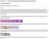
(This case study was added to OER Commons as one of a batch of over 700. It has relevant information which may include medical imagery, lab results, and history where relevant. A link to the final diagnosis can be found at the end of the case study for review. The first paragraph of the case study -- typically, but not always the clinical presentation -- is provided below.)
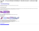
We present a case of plasma cell predominant lymphoplasmacyte-rich meningioma with numerous crystalline inclusions. A 72-year old woman presented with one-year history of memory disturbance and an MRI revealing a right frontal extra-axial mass. Histology showed a benign meningioma with multifocal accumulation of numerous large cells with abundant eosinophilic cytoplasm, filled with lamellar inclusions and bipyramidal crystals. In addition, some classic plasma cells, mastocytes and lymphocytes were also detected. The crystal containing cells were positive by CD79a and PGM1 and expressed polyclonal light chains, with kappa predominance. Electron microscopy showed crystals with needle or rectangular shape in plasma cells. Plasma cell inclusions have been reported occasionally in reactive inflammatory lesions but more frequently in plasma cell tumors and lymphoplasmacytic lymphoma, maybe associated with crystal-storing histiocytosis. Although plasma cell predominant lymphoplasmacyte-rich meningioma is not uncommon, to our knowledge, similar crystalline inclusions in plasma cells have not been reported previously in meningioma.
- Subject:
- Applied Science
- Education
- Health, Medicine and Nursing
- Life Science
- Material Type:
- Case Study
- Diagram/Illustration
- Provider:
- University of Pittsburgh School of Medicine
- Provider Set:
- Department of Pathology
- Author:
- Istvan Bodi
- Stefan Buk
- Tibor Hortobágyi
- Date Added:
- 08/01/2022
