Updating search results...

Search Resources

740 Results

View
Selected filters:
  • University of Pittsburgh
Pathology Case Study: A 9 year old girl with progressive weakness
Read the Fine Print
Some Rights Reserved
Rating
0.0 stars

(This case study was added to OER Commons as one of a batch of over 700. It has relevant information which may include medical imagery, lab results, and history where relevant. A link to the final diagnosis can be found at the end of the case study for review. The first paragraph of the case study -- typically, but not always the clinical presentation -- is provided below.)

A 9-year old girl presented with a 6 month history of progressive shoulder and back discomfort associated with generalized weakness and exercise intolerance.

Subject:
Applied Science
Education
Health, Medicine and Nursing
Life Science
Material Type:
Case Study
Diagram/Illustration
Provider:
University of Pittsburgh School of Medicine
Provider Set:
Department of Pathology
Author:
Chitra Prasad
Craig Campbell
Natashia Seemann
Robert Hammond
Date Added:
08/01/2022
Pathology Case Study: A 9-year-old girl with recurrent bilateral ovarian masses
Read the Fine Print
Some Rights Reserved
Rating
0.0 stars

(This case study was added to OER Commons as one of a batch of over 700. It has relevant information which may include medical imagery, lab results, and history where relevant. A link to the final diagnosis can be found at the end of the case study for review. The first paragraph of the case study -- typically, but not always the clinical presentation -- is provided below.)

A CT scan revealed multiloculated, multicystic masses arising from the pelvis to the level of the umbilicus and ovarian in origin. She underwent a laparoscopic decompression of the cysts together with biopsy of the cyst wall. The cytology was benign and the cyst wall was diagnosed as "luteinized follicular cysts" with a diagnostic note that since the cysts were large and bilateral, a non-ovarian endocrine effect was possible. After two months, the ovarian cysts recurred larger than before (CT scan, Figure 1) and were removed laparoscopically. These cysts were diagnosed as multiple luteinized cysts with prominent granulosa cell layer. Outside expert consultation was sought because of the clinical concern of their rapid recurrence. The expert consultant diagnosed the cysts as "multicystic juvenile granulosa cell tumors". The in-house diagnosis was amended accordingly. Four months after her initial presentation, a bilateral salpingo-oophorectomy was performed. Post surgery she was started on chemotherapy (Cisplatin, VP-16 and Bleomycin). At the time of her first chemotherapy administration she was noted on admission to have symptoms of clinical hypothyroidism including dry skin, obesity, cold intolerance, constipation and short stature.

Subject:
Applied Science
Education
Health, Medicine and Nursing
Life Science
Material Type:
Case Study
Diagram/Illustration
Provider:
University of Pittsburgh School of Medicine
Provider Set:
Department of Pathology
Author:
.BCh
Akosua B. DomfehChB
Ronald Jaffe
Date Added:
08/01/2022
Pathology Case Study: A Bioterror Agent in the Laboratory
Read the Fine Print
Some Rights Reserved
Rating
0.0 stars

(This case study was added to OER Commons as one of a batch of over 700. It has relevant information which may include medical imagery, lab results, and history where relevant. A link to the final diagnosis can be found at the end of the case study for review. The first paragraph of the case study -- typically, but not always the clinical presentation -- is provided below.)

A teenager presents to an outside emergency department with complaints of left eye pressure, itchiness, yellow drainage, left jaw pain and difficulty opening the mouth. The teenager was camping in Ohio. A computed topography was performed on the orbit which demonstrated preseptal cellulitis without abscess formation. The plan at that time was to be discharged home with polymyxin/trimethoprim ophthalmology drops and oral sulfamethoxazole/trimethoprim. The patient returned to the emergency department one day later with worsening of symptoms to include eye pain, redness, sore throat, neck pain, headache, fever (101 F), and surrounding swelling. The patient was admitted to the hospital for intravenous ceftriaxone and clindamycin. Blood cultures after two days were negative. At this point, the patient was transferred to a larger hospital. Laboratory values upon admission were as follows: white blood cell count (WBC) = 8.1, platelets (PLT) = 280, hemoglobin = 14.5, hematocrit = 41.1. erythrocyte sedimentation rate (ESR) = 15 and C-reactive protein (CRP) = 3.5. On physical exam there was significant left conjunctival injection, watering, and periorbital swelling. The extraocular movements were intact. Ophthalmology was consulted with the impression of hemorrhagic conjunctivitis commonly due to adenovirus. Ophthalmology recommended artificial tears, discontinue antibiotics and discharge home to follow up in clinic in one to two weeks. Two days later, the patient returned to the emergency department with a fever of 103 F. The patient had decreased activity and decreased intake of fluids. There was continued complaints of headaches, neck pain, sore throat, and discomfort in the left eye. Physical exam at this time demonstrated left eye pain, redness, swelling, and a swollen lymph node on the left side of the face. Laboratory values at this time were as follows: WBC = 16.1, PLT = 402, hemoglobin = 12.8, hematocrit = 37.2, ESR = 48 and CRP = 9.47. Ophthalmology was consulted again with a recommendation to consult infectious disease and send testing for Bartonella and Tularemia. The infectious disease team discovered that during the camping trip the patient handled a raccoon without gloves. Infectious disease recommendations were to take doxycycline and to send serologic testing for Lyme disease, Bartonella, Ehrlichia, and Tularemia. In addition, a conjunctival swab was taken for culture. Originally, the conjunctival swab was sent to a small microbiology laboratory. The specimen went through routine specimen processing which resulted in the isolation of a small Gram-negative rod which underwent Kirby Bauer susceptibility testing. No identification was made so the specimen was referred to a larger laboratory. At the larger laboratory the specimen did not grow on blood agar plate but demonstrated some growth on chocolate agar plate. A gram stain was performed on the culture which demonstrated small gram-negative rods. The presumptive identification was Haemophilus because of the gram stain pattern and the clinical history of an ocular source. The organism was examined by MALDI-TOF which did not identify the organism. At this point 16S sequencing was performed. The results of the 16S sequencing (Figure 1.) demonstrated a probable identification of Francisella tularensis. The isolate was forwarded to the State Health Department Laboratory who confirmed the identification by PCR and direct immunofluorescence. The patient followed up in clinic a week later with reports that the eye is much better with no complaints of pain, redness, tearing, discharge or blurry vision.
Subject:
Applied Science
Education
Health, Medicine and Nursing
Life Science
Material Type:
Case Study
Diagram/Illustration
Provider:
University of Pittsburgh School of Medicine
Provider Set:
Department of Pathology
Author:
. William Pasculle
Jacob A Smith
ScD.
Date Added:
08/01/2022
Pathology Case Study: A Child with Radiologically Recurrent Thalamic Tumor
Read the Fine Print
Some Rights Reserved
Rating
0.0 stars

(This case study was added to OER Commons as one of a batch of over 700. It has relevant information which may include medical imagery, lab results, and history where relevant. A link to the final diagnosis can be found at the end of the case study for review. The first paragraph of the case study -- typically, but not always the clinical presentation -- is provided below.)

A 3-year-old boy initially presented in January 2010, with a 4-month history of progressive right hemiparesis. Developmentally his motor skills had been somewhat delayed, with walking only at 18 months while dragging his right leg and a left hand preference from an early age. Initial MRI revealed a large left thalamic enhancing partly cystic-solid lesion involving the left posterior limb of the internal capsule and the posterior lentiform, with mass effect on the third ventricle and brainstem (Figure 1, T1 post contrast) (Figure 2, T2) (Figure 3, FLAIR). MRI of the spine was normal.

Subject:
Applied Science
Education
Health, Medicine and Nursing
Life Science
Material Type:
Case Study
Diagram/Illustration
Provider:
University of Pittsburgh School of Medicine
Provider Set:
Department of Pathology
Author:
Chandrasekaran Kaliaperumal
Elizabeth Ryan
Francesca Brett
John Caird
Linda Mulligan
Seamus Looby
Tafadzwa Mandiwanza
Date Added:
08/01/2022
Pathology Case Study: A Female Child with 21q Deletion
Read the Fine Print
Some Rights Reserved
Rating
0.0 stars

(This case study was added to OER Commons as one of a batch of over 700. It has relevant information which may include medical imagery, lab results, and history where relevant. A link to the final diagnosis can be found at the end of the case study for review. The first paragraph of the case study -- typically, but not always the clinical presentation -- is provided below.)

The patient is a 15-month-old female who was born to a GP2A4 healthy mother following an uncomplicated antenatal course. The birth was complicated by shoulder dystocia and tight nuchal cord. Following birth, the patient required NICU admission for respiratory support. Additionally, the neonate showed oozing of blood, hypotonia, possible seizures and lack of feeding cues or interest. Investigations revealed thrombocytopenia (55,000/µL), multiple cardiac anomalies (including persistent pulmonary hypertension, perimembranous ventricular septal defect, secundum atrial septal defect and bidirectional patent ductus arteriosus), intracranial bleeding (including left parietal cephalohematoma, multiple subdural hematomas and findings suggestive of prior intraventricular hemorrhage) and central nervous system anomalies (extensive polymicrogyria involving the posterior cerebral hemispheres and simplified gyration in bilateral frontal lobes).

Subject:
Applied Science
Education
Health, Medicine and Nursing
Life Science
Material Type:
Case Study
Diagram/Illustration
Provider:
University of Pittsburgh School of Medicine
Provider Set:
Department of Pathology
Author:
Eric Carlsen
Grant Bullock
Marie DeFrances
Melanie Babcock
Shikha Malhotra
Svetlana Yatsenko
Date Added:
08/01/2022
Pathology Case Study: A Female Infant with Fever and Pancytopenia
Read the Fine Print
Some Rights Reserved
Rating
0.0 stars

(This case study was added to OER Commons as one of a batch of over 700. It has relevant information which may include medical imagery, lab results, and history where relevant. A link to the final diagnosis can be found at the end of the case study for review. The first paragraph of the case study -- typically, but not always the clinical presentation -- is provided below.)

A 3 month previously healthy female with no significant family history presented with a 2 day history of fever (38.8 °C) abdominal distention and tachypnea. Initial laboratory evaluation demonstrated pancytopenia with absolute neutropenia, elevated liver function tests, prolonged coagulation times, hyperlactatemia and elevated ferritin levels (Table 1). An infectious workup was negative (EBV, CMV, Adenovirus, Influenza, Parainfluenza, RSV, Chlamydia, Mycoplasma, Anaplasma and all cultures). A bone marrow biopsy was performed and demonstrated histiocytes with hemophagocytosis (Figure 1). Further testing demonstrated elevated soluble IL-2 receptor levels (>20000 pg/mL (Reference range: 622 to 1619 pg/mL). Subsequently genetic testing ordered was positive for biallelic mutations in PRF1.

Subject:
Applied Science
Education
Health, Medicine and Nursing
Life Science
Material Type:
Case Study
Diagram/Illustration
Provider:
University of Pittsburgh School of Medicine
Provider Set:
Department of Pathology
Author:
Daniel Martinez
Nidhi Aggarwal
Date Added:
08/01/2022
Pathology Case Study: A Female Patient in Her 50's with Shortness of Breath
Read the Fine Print
Some Rights Reserved
Rating
0.0 stars

(This case study was added to OER Commons as one of a batch of over 700. It has relevant information which may include medical imagery, lab results, and history where relevant. A link to the final diagnosis can be found at the end of the case study for review. The first paragraph of the case study -- typically, but not always the clinical presentation -- is provided below.)

The patient is a 54-year-old female presenting with shortness of breath, malaise, and cough.

Subject:
Applied Science
Education
Health, Medicine and Nursing
Life Science
Material Type:
Case Study
Diagram/Illustration
Provider:
University of Pittsburgh School of Medicine
Provider Set:
Department of Pathology
Author:
Daniel Martinez
Paul Ohori
Date Added:
08/01/2022
Pathology Case Study: A Female in Her 50s with  Asymptomatic Renal Mass
Read the Fine Print
Some Rights Reserved
Rating
0.0 stars

(This case study was added to OER Commons as one of a batch of over 700. It has relevant information which may include medical imagery, lab results, and history where relevant. A link to the final diagnosis can be found at the end of the case study for review. The first paragraph of the case study -- typically, but not always the clinical presentation -- is provided below.)

The patient is a female in her 50s who presents with asymptomatic, incidentally detected renal mass. Personal history is relevant for low-grade appendiceal mucinous neoplasm (LAMN, pTis), and family history of ovarian and gastric cancer. Smoking history is negative. CT scan and MRI of abdomen demonstrate a left lower renal pole enhancing lesion (2.0 cm). The patient elects to undergo a laparoscopic partial nephrectomy of the mass, instead of active surveillance or percutaneous biopsy/ cryoablation.

Subject:
Applied Science
Education
Health, Medicine and Nursing
Life Science
Material Type:
Case Study
Diagram/Illustration
Provider:
University of Pittsburgh School of Medicine
Provider Set:
Department of Pathology
Author:
Fawaz Almutairi
Gabriela Quiroga-Garza
Shikha Malhotra
Date Added:
08/01/2022
Pathology Case Study: A Female in Her 60's with  Diplopia
Read the Fine Print
Some Rights Reserved
Rating
0.0 stars

(This case study was added to OER Commons as one of a batch of over 700. It has relevant information which may include medical imagery, lab results, and history where relevant. A link to the final diagnosis can be found at the end of the case study for review. The first paragraph of the case study -- typically, but not always the clinical presentation -- is provided below.)

The patient is a 60-70 year old female with a history of increased intraocular pressure felt to be due to ocular hypertension versus glaucoma and a selective laser trabeculoplasty of her left eye. She presented to her ophthalmologist for a routine intraocular pressure check. On examination, she was noted to have diplopia, proptosis, and limitation of elevation/abduction and no pain with orbital movement. A CT sinus/orbit was performed which showed an ill- defined mass within the left retrobulbar, superonasal soft tissue and involving the left lacrimal gland (see Fig 1). A lacrimal gland biopsy was performed which showed mild chronic dacryoadenitis, but ophthalmology was concerned that the mass was not adequately sampled and proceeded with a retrobulbar biopsy. This biopsy was received by Hematopathology in consultation without accompanying flow cytometric immunophenotypic studies.

Subject:
Applied Science
Education
Health, Medicine and Nursing
Life Science
Material Type:
Case Study
Diagram/Illustration
Provider:
University of Pittsburgh School of Medicine
Provider Set:
Department of Pathology
Author:
Nidhi Aggarwal
Terri Jones
Date Added:
08/01/2022
Pathology Case Study: A Female in Her 60s with a Kidney Mass
Read the Fine Print
Some Rights Reserved
Rating
0.0 stars

(This case study was added to OER Commons as one of a batch of over 700. It has relevant information which may include medical imagery, lab results, and history where relevant. A link to the final diagnosis can be found at the end of the case study for review. The first paragraph of the case study -- typically, but not always the clinical presentation -- is provided below.)

The patient is a female in her 60s who presents with an incidentally detected kidney mass. Computed tomography shows an enhancing 2.0 cm exophytic, solid right kidney mass of the lower pole. She denies any symptoms, smoking history, or family history of urological malignancies. The patient elects to undergo a laparoscopic partial nephrectomy of the mass in lieu of continued surveillance.

Subject:
Applied Science
Education
Health, Medicine and Nursing
Life Science
Material Type:
Case Study
Diagram/Illustration
Provider:
University of Pittsburgh School of Medicine
Provider Set:
Department of Pathology
Author:
Daniel Geisler
Gabriela Quiroga-Garza
Date Added:
08/01/2022
Pathology Case Study: A Female in Her 70s Presenting with a Breast Mass
Read the Fine Print
Some Rights Reserved
Rating
0.0 stars

(This case study was added to OER Commons as one of a batch of over 700. It has relevant information which may include medical imagery, lab results, and history where relevant. A link to the final diagnosis can be found at the end of the case study for review. The first paragraph of the case study -- typically, but not always the clinical presentation -- is provided below.)

A female in her early 70s was found to have a left breast lesion (detected on imaging studies) at an outside institution. It measured 0.7 cm and was located in the upper outer quadrant at 1 O'clock position. Dermatopathology services received the slides as a consult with limited history. The case was subsequently forwarded to division of breast pathology for further characterization.

Subject:
Applied Science
Education
Health, Medicine and Nursing
Life Science
Material Type:
Case Study
Diagram/Illustration
Provider:
University of Pittsburgh School of Medicine
Provider Set:
Department of Pathology
Author:
Rohit Bhargava
Sonal Choudhary
Vandana Baloda
Date Added:
08/01/2022
Pathology Case Study: A Female in Her 80s with a Renal Mass
Read the Fine Print
Some Rights Reserved
Rating
0.0 stars

(This case study was added to OER Commons as one of a batch of over 700. It has relevant information which may include medical imagery, lab results, and history where relevant. A link to the final diagnosis can be found at the end of the case study for review. The first paragraph of the case study -- typically, but not always the clinical presentation -- is provided below.)

An 80 plus year-old female with past medical history of coronary disease was found to have an incidental mass in the right kidney during an ultrasound performed for a recent elevated creatinine level. No previous history of chronic renal failure, hypertension or diabetes was reported.

Subject:
Applied Science
Education
Health, Medicine and Nursing
Life Science
Material Type:
Case Study
Diagram/Illustration
Provider:
University of Pittsburgh School of Medicine
Provider Set:
Department of Pathology
Author:
Angela Sanguino Ramirez
Rajiv Dhir
Thu Tran
Date Added:
08/01/2022
Pathology Case Study: A Female in Her Early 60s with Worsened Joint Pain after Initiation of Immunotherapy
Read the Fine Print
Some Rights Reserved
Rating
0.0 stars

(This case study was added to OER Commons as one of a batch of over 700. It has relevant information which may include medical imagery, lab results, and history where relevant. A link to the final diagnosis can be found at the end of the case study for review. The first paragraph of the case study -- typically, but not always the clinical presentation -- is provided below.)

The patient is a 61-year-old female with a 40-pack-year history of smoking and KRAS mutation positive stage IV lung adenocarcinoma with osseous involvement and initial response to four cycles of traditional chemotherapy. She then experienced disease progression and was started on palliative radiotherapy and second-line treatment with the checkpoint inhibitor nivolumab (Opdivo).

Subject:
Applied Science
Education
Health, Medicine and Nursing
Life Science
Material Type:
Case Study
Diagram/Illustration
Provider:
University of Pittsburgh School of Medicine
Provider Set:
Department of Pathology
Author:
Jenn Yoest
Date Added:
08/01/2022
Pathology Case Study: A Female with Chief Complaints of Fever, Productive Cough, Night Sweats and Weight Loss
Read the Fine Print
Some Rights Reserved
Rating
0.0 stars

(This case study was added to OER Commons as one of a batch of over 700. It has relevant information which may include medical imagery, lab results, and history where relevant. A link to the final diagnosis can be found at the end of the case study for review. The first paragraph of the case study -- typically, but not always the clinical presentation -- is provided below.)

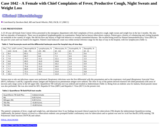
A 25-30 year old female from Central Africa presented to the emergency department with chief complaints of fever, productive cough, night sweats and weight loss in the last 2 months. She also had two episodes of hemoptysis. There was no peripheral lymphadenopathy on examination. Patient had no known tuberculosis contacts. Patient gave a history of volunteering and visiting hospitals on weekends in her country of origin. She did not have any history of high-risk behavior or sexually transmitted diseases. She recalled being tested for Human Immunodeificiency Virus (HIV) in the past few years and was found to be negative. Patient's total leukocyte count was within normal reference range for the days of stay in the hospital, with low lymphocytes (Table 1).

Subject:
Applied Science
Education
Health, Medicine and Nursing
Life Science
Material Type:
Case Study
Diagram/Illustration
Provider:
University of Pittsburgh School of Medicine
Provider Set:
Department of Pathology
Author:
Davsheen Bedi
Sarah Wheeler
Date Added:
08/01/2022
Pathology Case Study: A Female with an Anti-Microbial Refractory Fungal Infection
Read the Fine Print
Some Rights Reserved
Rating
0.0 stars

(This case study was added to OER Commons as one of a batch of over 700. It has relevant information which may include medical imagery, lab results, and history where relevant. A link to the final diagnosis can be found at the end of the case study for review. The first paragraph of the case study -- typically, but not always the clinical presentation -- is provided below.)

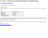
A woman in her 60s with a history of relapsed AML status-post matched unrelated donor allogenic stem cell transplant continued to have pancytopenia with evidence of poor engraftment (lab values in Table 1. below).

Subject:
Applied Science
Education
Health, Medicine and Nursing
Life Science
Material Type:
Case Study
Diagram/Illustration
Provider:
University of Pittsburgh School of Medicine
Provider Set:
Department of Pathology
Author:
Darrell Triulzi
Ian M. Harrold
Date Added:
08/01/2022
Pathology Case Study: A Frontal Dural-Based Lesion in a 63-Year Old Male
Read the Fine Print
Some Rights Reserved
Rating
0.0 stars

(This case study was added to OER Commons as one of a batch of over 700. It has relevant information which may include medical imagery, lab results, and history where relevant. A link to the final diagnosis can be found at the end of the case study for review. The first paragraph of the case study -- typically, but not always the clinical presentation -- is provided below.)

A 63-year old male presented to a neurosurgeon with a firm, immobile, non-tender mass of his right supraorbital forehead. The patient endorsed remote (x 40 years) trauma to the area with subsequent bony irregularity, with recent (x 3 months) subtle enlargement. There was no associated neurologic symptom and his physical exam was negative for neurologic deficits. Imaging revealed a 3.9-centimeter dural-based lesion with osseous extension involving both inner and outer table; there was a dural tail with enhancement, but not bulky intracranial extension. A frontal craniotomy with tumor resection including wide dural excision was performed at an outside institution. The pathologic diagnosis rendered was meningioma; there was disagreement regarding whether to classify the neoplasm as 'atypical'. No adjuvant therapy was administered. Three years later the lesion recurred in the midline, adjacent to the resection site (Figure 1A, Axial T1-weighted MRI with contrast enhancement), and the patient presented to Moffitt Cancer Center for further neurosurgical management. The tumor was resected and the diagnosis was unchanged at that time. The tumor recurred and was re-excised two years later, prompting re-evaluation of the case. A fourth operation was undertaken the following year, six years after the initial presentation; histomorphology was unchanged.

Subject:
Applied Science
Education
Health, Medicine and Nursing
Life Science
Material Type:
Case Study
Diagram/Illustration
Provider:
University of Pittsburgh School of Medicine
Provider Set:
Department of Pathology
Author:
Arnold B. Etame
Ashley R. Perkins
Julia A. Bridge
Marilyn M. Bui
Robert J.B. Macaulay
Date Added:
08/01/2022
Pathology Case Study: A Gentleman with a Lump in his Jaw
Read the Fine Print
Some Rights Reserved
Rating
0.0 stars

(This case study was added to OER Commons as one of a batch of over 700. It has relevant information which may include medical imagery, lab results, and history where relevant. A link to the final diagnosis can be found at the end of the case study for review. The first paragraph of the case study -- typically, but not always the clinical presentation -- is provided below.)

At follow-up with his primary care physician two weeks later, the mass had not decreased in size. The primary care physician was concerned for malignancy, particularly given the patient's history of smoking and alcohol use, and a CT neck was performed. CT revealed a 2.3 x 1.8 cm soft tissue mass in the right neck with surrounding inflammation (performed at an outside institution; images not available). At this point the gentleman was referred to ENT with concern for possible carcinoma, and an incisional biopsy was performed. Tissue was sent for surgical pathology and aerobic culture.

Subject:
Applied Science
Education
Health, Medicine and Nursing
Life Science
Material Type:
Case Study
Diagram/Illustration
Provider:
University of Pittsburgh School of Medicine
Provider Set:
Department of Pathology
Author:
A. William Pasculle
Sarah B. Hugar
ScD.
Date Added:
08/01/2022
Pathology Case Study: A Male Newborn with Bruising and Petechia
Read the Fine Print
Some Rights Reserved
Rating
0.0 stars

(This case study was added to OER Commons as one of a batch of over 700. It has relevant information which may include medical imagery, lab results, and history where relevant. A link to the final diagnosis can be found at the end of the case study for review. The first paragraph of the case study -- typically, but not always the clinical presentation -- is provided below.)

A male newborn was delivered at gestational age 37 weeks and 1 day via spontaneous vaginal delivery. His teenage mother was G1P1 and received routine antenatal care. His mother's blood type was A- (for which Rhogam treatment was received) and her pregnancy was complicated by preeclampsia.

Subject:
Applied Science
Education
Health, Medicine and Nursing
Life Science
Material Type:
Case Study
Diagram/Illustration
Provider:
University of Pittsburgh School of Medicine
Provider Set:
Department of Pathology
Author:
Alesia Kaplan
Dayne Ashman
Date Added:
08/01/2022
Pathology Case Study: A Male Undergoing Therapeutic Drug Monitoring for Busulfan Dosing
Read the Fine Print
Some Rights Reserved
Rating
0.0 stars

(This case study was added to OER Commons as one of a batch of over 700. It has relevant information which may include medical imagery, lab results, and history where relevant. A link to the final diagnosis can be found at the end of the case study for review. The first paragraph of the case study -- typically, but not always the clinical presentation -- is provided below.)

The patient is a male is his early 60s with a history of primary refractory AML who was admitted to the hospital for matched unrelated bone marrow transplantation. He was on prophylactic metronidazole before admission, and unbeknownst to the clinical team, he took a dose of metronidazole the evening before beginning induction with Flu/Bu4/ATG. Per protocol at our institution, therapeutic drug monitoring (TDM) of busulfan was performed by collecting blood samples at time zero and at 3, 4, 6 and 8 hours after beginning the infusion. The drug levels were measured using HPLC-MS/MS. The values obtained for all four busulfan doses on this patient are graphed in Figure 1 and presented in Table 1. The target for average busulfan area under the curve (AUC) was 4800 uMol/L*min for four doses, given every 24 hours for 4 days. With this in mind, the first dose given was 240 mg. However, when the busulfan levels were measured after the first dose, the AUC was 7372 uMol/L*min, which represents 154% of the expected level. The clearance rate was calculated to be 1.93 mL/min/kg, but is expected to be 3.0 mL/min/kg. At this point, the clinical team became aware that the patient had taken the metronidazole dose. Additionally, for the desired average AUC target of 4800 uMol/L*min, a -47% dose adjustment, for a dose of 128 mg, was recommended to begin at dose 2. TDM for the second dose showed an AUC of 3920 uMol/L*min. The clearance was 1.94 mL/min/kg, which is very similar to the first dose, indicating the effects of metronidazole were ongoing. For day 3, a dose of 128 mg was also used, and this time the AUC was 3184 uMol/L*min with a clearance of 2.39 mL/min/kg. This increased clearance rate indicated the effects of metronidazole were beginning to wear off, and a higher dose of 190 mg was recommended for the fourth dose of busulfan. TDM for the fourth dose showed an AUC of 4720 uMol/L*min and a clearance rate again of 2.39 mL/min/kg. The average AUC for the four doses was 4799 uMol/L*min, which was very close to the target. A clearance rate of 2.39 mL/min/kg, however, is still below the anticipated clearance of 3.0 mL/min/kg, suggesting that even at approximately 80 hours after metronidazole ingestion, it was still effecting the busulfan clearance.

Subject:
Applied Science
Education
Health, Medicine and Nursing
Life Science
Material Type:
Case Study
Diagram/Illustration
Provider:
University of Pittsburgh School of Medicine
Provider Set:
Department of Pathology
Author:
Megan Zilla
Raman Venkataramanan
Date Added:
08/01/2022
Pathology Case Study: A Male in His 50s with Urothelial Carcinoma
Read the Fine Print
Some Rights Reserved
Rating
0.0 stars

(This case study was added to OER Commons as one of a batch of over 700. It has relevant information which may include medical imagery, lab results, and history where relevant. A link to the final diagnosis can be found at the end of the case study for review. The first paragraph of the case study -- typically, but not always the clinical presentation -- is provided below.)

The patient is a man in his fifties with an incidental finding of a 5 cm enhancing renal mass on the right upper pole during low-dose screening CT scan performed for history of tobacco use. He denies flank pain, gross hematuria, fever, chills, nausea, vomiting, night sweats, and change in appetite or weight loss. He is not aware of any family history of renal cancer. The patient's CT scan of the abdomen/pelvis exhibits a lesion in the upper pole of the right kidney highly concerning for a hypo-enhancing solid renal tumor/carcinoma, an additional simple cortical cyst in the right kidney, and one cortical cystic lesion in the lower pole of the left kidney, too small to further characterize. There is no evidence of adenopathy or metastatic disease.

Subject:
Applied Science
Education
Health, Medicine and Nursing
Life Science
Material Type:
Case Study
Diagram/Illustration
Provider:
University of Pittsburgh School of Medicine
Provider Set:
Department of Pathology
Author:
Dimitrios Korentzelos
Gabriela M Quiroga-Garza
Date Added:
08/01/2022