(This case study was added to OER Commons as one of a batch of over 700. It has relevant information which may include medical imagery, lab results, and history where relevant. A link to the final diagnosis can be found at the end of the case study for review. The first paragraph of the case study -- typically, but not always the clinical presentation -- is provided below.)
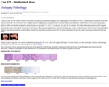
A male in his 30s with a long history of epilepsy was evaluated for a seizure. A general neurologic examination was unremarkable. Radiologic studies of the brain revealed a right frontal mass. Multiplanar, multisequential magnetic resonance images of the brain with intravenous gadolinium showed a homogeneously enhancing subfrontal extra-axial mass measuring 3.5 x 2.3 x 2.0 cm located right to the midline with surrounding edema, minimal midline shift and mild deformity of the right frontal horn (Fig. 1A). The lesion was broad based with dural extension into the anterior falx. Sagittal images showed irregular margins at the brain interface suggesting an intra-axial component (Fig. 1B). At the time of surgery, the frontal cortex was noted to have a "rock" hard consistency. Both the cortical and extra-axial (dural) components were grossly completely excised.
- Subject:
- Applied Science
- Education
- Health, Medicine and Nursing
- Life Science
- Material Type:
- Case Study
- Diagram/Illustration
- Provider:
- University of Pittsburgh School of Medicine
- Provider Set:
- Department of Pathology
- Author:
- Iezza G1
- Lanman TH3
- Loh C2
- Yong WH1
- Date Added:
- 08/01/2022
