(This case study was added to OER Commons as one of a …
(This case study was added to OER Commons as one of a batch of over 700. It has relevant information which may include medical imagery, lab results, and history where relevant. A link to the final diagnosis can be found at the end of the case study for review. The first paragraph of the case study -- typically, but not always the clinical presentation -- is provided below.)
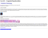
The patient, a 14-year-old with hypothyroidism since birth for which he was taking Synthroid (Levothyroxine), developed a nodule in the right lobe of the thyroid. The clinical working diagnosis was autoimmune thyroiditis. The family history was non-contributory. An ultrasound scan showed a hypoechoic nodule, and the thyroid function tests were all within normal limits. A fine needle aspiration was non-diagnostic; hence a right thyroid lobectomy was performed.
(This case study was added to OER Commons as one of a …
(This case study was added to OER Commons as one of a batch of over 700. It has relevant information which may include medical imagery, lab results, and history where relevant. A link to the final diagnosis can be found at the end of the case study for review. The first paragraph of the case study -- typically, but not always the clinical presentation -- is provided below.)
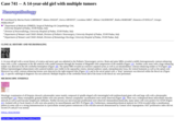
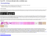
A 14-year-old girl with a recent history of sciatica and ataxic gait was admitted to the Pediatric Neurosurgery service. Brain and spine MRIs revealed a mildly heterogeneously contrast-enhancing mass with a cystic component in the 4th ventricle with caudal extension through the foramen of Magendie with compression of the medulla (Figure 1A). Another cystic mass with a large enhancing nodule was detected in the left cerebellar hemisphere (Figure 1B). The spinal MRI revealed an extensive septated syrinx as well as an intramedullary contrast-enhancing nodule at T4 (Figure 1D). Diffuse leptomeningeal enhancement with scattered nodules was visible in the suprasellar cistern, internal auditory canals, interpeduncular fossa, the ventral brainstem as well as the spinal cord. Moreover two solid, contrast-enhancing lesions were detected in the right temporal lobe (Figure 1C); and in the sacral spinal cord, a "drop" metastasis was detected within the thecal sac (Figure 1E). A specific radiological diagnosis was not achieved. Multiple biopsies of the cerebellar lesion and of the tissue in the thecal sac were performed.
(This case study was added to OER Commons as one of a …
(This case study was added to OER Commons as one of a batch of over 700. It has relevant information which may include medical imagery, lab results, and history where relevant. A link to the final diagnosis can be found at the end of the case study for review. The first paragraph of the case study -- typically, but not always the clinical presentation -- is provided below.)
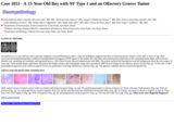
Our patient is a 15 year-old boy, with a previous diagnosis of neurofibromatosis type 2, who had undergone surgical resections of olfactory groove tumors, at five and 13 years of age. Both resections had histopathologically-confirmed meningothelial meningioma (WHO grade I).The patient also had other intracranial lesions bilaterally in the cerebellopontine angle, and in the left Mackel cave, involving the vestibular and trigeminal nerves. All of these lesions showed enhancement with MRI. The patient suffered left hearing loss and left hemiparesis since the first surgery 10 years ago. Three months before admission, the patient developed seizures and antiepileptic drugs were introduced. MRI showed relapse in the olfactory groove lesion (Fig. 1a). At this time, a third microsurgical approach of the olfactory groove lesion was performed, achieving satisfactory resection (Fig. 1b). The patient's condition did not worsen postoperatively.
(This case study was added to OER Commons as one of a …
(This case study was added to OER Commons as one of a batch of over 700. It has relevant information which may include medical imagery, lab results, and history where relevant. A link to the final diagnosis can be found at the end of the case study for review. The first paragraph of the case study -- typically, but not always the clinical presentation -- is provided below.)
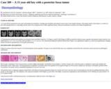
A 15 year old boy presented to our center with progressive headache, vomiting, gait instability and reduced visual acuity of one month's duration. The patient related a head injury as a result of a fall while playing hockey as coinciding with the onset of his symptoms but no other significant history. Physical examination revealed severe bilateral papilledema.
(This case study was added to OER Commons as one of a …
(This case study was added to OER Commons as one of a batch of over 700. It has relevant information which may include medical imagery, lab results, and history where relevant. A link to the final diagnosis can be found at the end of the case study for review. The first paragraph of the case study -- typically, but not always the clinical presentation -- is provided below.)
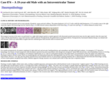
A 15 year-old female presented to the Emergency Room with urinary retention and inability to walk. She had developed progressive low back pain and bilateral leg pain about 2 months prior to presentation. Physical examination demonstrated bilateral positive Babinski reflex, bilateral positive Hoffmann's reflex, positive Romberg reflex and positive bulbocavernous reflex. Sagittal T1 weighted MRI scan post Gadolinium (Gd) using fat saturation technique showed an intensely enhancing intraspinal, extradural, 6.5 X 0.8 X 2.3 cm (cranial-caudal, anterior-posterior, transverse) mass, with anterior displacement and compression of the spinal cord (Figure 1A). The mass extended from T3 to T7. Axial T1 weighted MRI scan post Gd at T5 showed bilateral transverse process and spinous process involvement (Figure 1B). Axial T1 weighted MRI scan after Gd showed enhancing tumor in the spinal canal and neural foramina from T4 to T7 (Figure 1C, scan was taken at T5). A decompression surgery was performed.
(This case study was added to OER Commons as one of a …
(This case study was added to OER Commons as one of a batch of over 700. It has relevant information which may include medical imagery, lab results, and history where relevant. A link to the final diagnosis can be found at the end of the case study for review. The first paragraph of the case study -- typically, but not always the clinical presentation -- is provided below.)
A 15-year-old girl presented to our institution with headache, vomiting, and nausea of 3-week duration. Physical examination showed no focal neurologic deficits. The patient's past medical, surgical, and family histories were all noncontributory.
(This case study was added to OER Commons as one of a …
(This case study was added to OER Commons as one of a batch of over 700. It has relevant information which may include medical imagery, lab results, and history where relevant. A link to the final diagnosis can be found at the end of the case study for review. The first paragraph of the case study -- typically, but not always the clinical presentation -- is provided below.)
The patient is a 15-year-old adolescent male with a history of developmental delay who presented at 8 to 10 years of age with loss of peripheral vision and night blindness, at which time he was diagnosed with retinitis pigmentosa. He then developed progressive loss of central vision and macular edema. He was evaluated by an ophthalmologist, subspecialzing in genetics. A diagnostic laboratory test was requested because of the atypical presentation of retinitis pigmentosa.
(This case study was added to OER Commons as one of a …
(This case study was added to OER Commons as one of a batch of over 700. It has relevant information which may include medical imagery, lab results, and history where relevant. A link to the final diagnosis can be found at the end of the case study for review. The first paragraph of the case study -- typically, but not always the clinical presentation -- is provided below.)
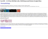
A 16 month old female was admitted to the Pediatric Intensive Care Unit with fever, hypoxia, altered mental status, and seizures. Magnetic resonance imaging (MRI) of the brain (Figure 1) showed a single well circumscribed intraventricular mass which was isointense to cortex on T1-and T2-weighted images, and demonstrated mildly restricted diffusion consistent with dense cell packing. The mass measured 4x4x4 cm in the craniocaudal, AP and transverse dimensions. FLAIR imaging revealed mass effect secondary to the tumor causing trapping of the left temporal horn. There was additional, but mild right lateral ventricular dilatation and an 11 mm midline shift as measured at the level of the anterior portion of the third ventricle. Edema was seen along the corpus callosum, surrounding the mass extending into the left occipital lobe, temporal lobe, and parietal lobe. These signal characteristics were most suggestive of an intraventricular meningioma or a supratentorial primitive neuroectodermal tumor.
(This case study was added to OER Commons as one of a …
(This case study was added to OER Commons as one of a batch of over 700. It has relevant information which may include medical imagery, lab results, and history where relevant. A link to the final diagnosis can be found at the end of the case study for review. The first paragraph of the case study -- typically, but not always the clinical presentation -- is provided below.)
A 16-year-old right-handed male presented to an emergency room after suffering mild head trauma from a fall onto his head while intoxicated with alcohol. Physical examination revealed a healthy, inebriated male with no focal neurological deficit. The mother reported that her son had experienced excessive fatigue over the last three months. The patient's past medical, surgical and family history were all noncontributory.
(This case study was added to OER Commons as one of a …
(This case study was added to OER Commons as one of a batch of over 700. It has relevant information which may include medical imagery, lab results, and history where relevant. A link to the final diagnosis can be found at the end of the case study for review. The first paragraph of the case study -- typically, but not always the clinical presentation -- is provided below.)
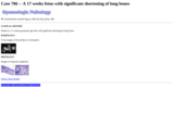
Patient is a 17 weeks gestational age fetus with significant shortening of long bones.
(This case study was added to OER Commons as one of a …
(This case study was added to OER Commons as one of a batch of over 700. It has relevant information which may include medical imagery, lab results, and history where relevant. A link to the final diagnosis can be found at the end of the case study for review. The first paragraph of the case study -- typically, but not always the clinical presentation -- is provided below.)
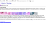
The patient is a 17-year-old female with a history of neurofibromatosis type 1 (NF1) and Arnold-Chiari type I malformation, who presented with painful enlargement of a subcutaneous left thigh mass. The mass, which had been present for several years, was previously stable at 1.5 cm and assumed to be a neurofibroma. Over several months, the lesion grew to 2.5 cm and became painful. These changes were worrisome for transformation of the assumed neurofibroma to a malignant nerve sheath tumor.
(This case study was added to OER Commons as one of a …
(This case study was added to OER Commons as one of a batch of over 700. It has relevant information which may include medical imagery, lab results, and history where relevant. A link to the final diagnosis can be found at the end of the case study for review. The first paragraph of the case study -- typically, but not always the clinical presentation -- is provided below.)
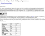
The patient is a 17 year-old previously healthy girl who developed Raynaud's phenomenon in late 2006. In early 2007, she developed fatigue and general malaise with reportedly increased ESR and CPK. In February, she had progressing symptoms, with periods of chills followed by sweating thought to be fevers that were never measured. In early March, she had joint pain, specifically in her hands, wrists, knees and ankles, as well as myalgias in her back and lower legs. In late March, she was admitted for appendicitis and had an appendectomy at which time an abdominal CT scan noted interstitial changes at the base of her lungs. In April, she was being followed as an outpatient by Rheumatology and Pulmonary Medicine at an outside hospital. She was found to have a positive rheumatoid factor and diagnosed with rheumatoid arthritis with possible associated pulmonary fibrosis. She was started on NSAIDS. Later that month, she began to have more joint pain and weakness. In early May, she was started on prednisone which was increased to 60 mg bid when she noticed significant improvement. In mid May, she began to have shortness of breath, which progressively worsened.
(This case study was added to OER Commons as one of a …
(This case study was added to OER Commons as one of a batch of over 700. It has relevant information which may include medical imagery, lab results, and history where relevant. A link to the final diagnosis can be found at the end of the case study for review. The first paragraph of the case study -- typically, but not always the clinical presentation -- is provided below.)
A 17-year-old girl presented with a seizure which started as involuntary movement of left arm and progressed to loss of consciousness. She experienced severe headaches for three weeks prior to hospital admission. There was no previous medical history. On neurologic examination, bilateral papilledema was noted and no other neurologic deficits were detected. MRI revealed a 7x4cm heterogeneous extra-axial mass in right frontoparietal area with no vasogenic edema. The mass contained a septated cystic portion with high signal intensity on T2 and low signal intensity on T1-weighted images. The solid portion had slightly higher signal intensity on both T1 and T2-weighted images. With gadolinium enhancement, the solid portion and the cystic wall showed strong enhancement. Perfusion was elevated in the solid portion (Figures 1, 2 and 3).
(This case study was added to OER Commons as one of a …
(This case study was added to OER Commons as one of a batch of over 700. It has relevant information which may include medical imagery, lab results, and history where relevant. A link to the final diagnosis can be found at the end of the case study for review. The first paragraph of the case study -- typically, but not always the clinical presentation -- is provided below.)
A 19-year-old male was referred for a blurred vision and eyestrain associated with occipital headache for several weeks. Ophthalmologic examination showed left homonymous hemianopia and papilledema on fundoscopy. The CT scan showed a 7 cm diameter lobulated hyperdense mass with calcifications. On MRI, the lesion was hyperintense on T2 and hypointense on T1 weighted images with moderate gadolinium enhancement. The lesion was well demarcated and laid on tentorium. Diffusion weighted imaging did not exhibit restriction (High apparent diffusion coefficient (ADC)). Surrounding brain parenchyma was normal (Figure 1). The lesion was hypoperfused when compared to normal brain (Figure 2). MRI spectroscopy (Figure 3) showed a high choline to creatine ratio (suggesting high cell membrane turn over) and elevated lipids and NAA. The lesion was totally removed via a right parietal craniotomy. Intraoperatively, the lesion was readily visible at the surface of the brain, firm, with a clear dissection plan. It was inserted on the falx cerebri and the tentorium. Gross examination showed a firm lobulated white lesion (Figure 4). There was no evidence of hemorrhage or necrosis.
(This case study was added to OER Commons as one of a …
(This case study was added to OER Commons as one of a batch of over 700. It has relevant information which may include medical imagery, lab results, and history where relevant. A link to the final diagnosis can be found at the end of the case study for review. The first paragraph of the case study -- typically, but not always the clinical presentation -- is provided below.)
A 19-year-old male with history of narcolepsy, but otherwise healthy with normal development and cognition, presented with one month of daily headache and a single unprovoked transient confusional episode consistent with a seizure. During the episode, the patient experienced right upper extremity incoordination, orolingual automatisms and aphasia. Physical examination was notable only for macrocephaly. MRI of the brain revealed multiple heterogeneously enhancing dural-based masses and dural nodularity with mild parenchymal volume loss, thinning and remodeling of the calvarium, remodeling of the skull base, and sagging appearance of brainstem (Figures 1a, 1b). There was no lesion in the spinal canal. Cerebrospinal fluid analysis was normal except for elevated protein content. Electroencephalography (EEG) showed left temporal focal slowing with sharp transients. Extensive serologic testing was within normal limits, notable for normal ANA, ANCA, RF, RPR, Quantiferon Gold, FSH, LH, prolactin, TSH, SPEP, antigliadin antibody, and IgG4, as well as negative HIV. CT scans of the chest, abdomen, and pelvis did not identify any visceral lesions and ophthalmologic and dermatologic examinations were essentially normal. A biopsy of the left parietal dural-based nodule was performed, but did not yield a definitive diagnosis. The patient was treated with levetiracetam and corticosteroid therapy and discharged home with planned outpatient follow up. Approximately four weeks later, he presented with recurrence of severe retro-orbital headache and emesis. A second biopsy, this time of a left frontal dural-based nodule was performed.
(This case study was added to OER Commons as one of a …
(This case study was added to OER Commons as one of a batch of over 700. It has relevant information which may include medical imagery, lab results, and history where relevant. A link to the final diagnosis can be found at the end of the case study for review. The first paragraph of the case study -- typically, but not always the clinical presentation -- is provided below.)
A 19-year-old male presented with an acute episode of headache, nausea and neck stiffness. The patient displayed a GCS of 15 with a mild left-sided hemiparesis. CCT revealed a mass in the right ventricular trigone with hemorrhage into the ventricle (Fig 1). An MRI showed contrast enhancement of the lesion (Fig 2; T2 axial view and Fig 3; T1 contrast-enhanced coronal view).
(This case study was added to OER Commons as one of a …
(This case study was added to OER Commons as one of a batch of over 700. It has relevant information which may include medical imagery, lab results, and history where relevant. A link to the final diagnosis can be found at the end of the case study for review. The first paragraph of the case study -- typically, but not always the clinical presentation -- is provided below.)
The patient is a 19-year-old female who presented to the emergency department (ED) with a two week history of general malaise, shaking chills, rash, fevers, night sweats and right upper quadrant pain. She had initially been seen two days prior in the ED, and preliminary evaluation demonstrated tender lymphadenopathy, an erythematous maculopapular rash of the malar region of the face, upper and lower extremities, and an elevated white blood cell count of 33,600 plt/L, with 5% "atypical cells" present. Her hemoglobin and hematocrit were within normal limits (14 g/dL and 42.2%), with an MCV of 84.9. At the time she was deemed well enough to follow up as outpatient with dermatology the following morning; by the time of this appointment she had developed shaking chills. This, in conjunction with the findings of the peripheral blood smear, prompted a second evaluation in the ED, with consultation by hematology
(This case study was added to OER Commons as one of a …
(This case study was added to OER Commons as one of a batch of over 700. It has relevant information which may include medical imagery, lab results, and history where relevant. A link to the final diagnosis can be found at the end of the case study for review. The first paragraph of the case study -- typically, but not always the clinical presentation -- is provided below.)
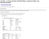
The patient is a 19-year-old male with a two-week history of febrile illness, respiratory failure, and septic shock. His illness started with low grade fever, intermittent headache, and nausea. Gradually, his symptoms progressed into high fever, prominent weakness, shortness of breath, and respiratory failure.
(This case study was added to OER Commons as one of a …
(This case study was added to OER Commons as one of a batch of over 700. It has relevant information which may include medical imagery, lab results, and history where relevant. A link to the final diagnosis can be found at the end of the case study for review. The first paragraph of the case study -- typically, but not always the clinical presentation -- is provided below.)
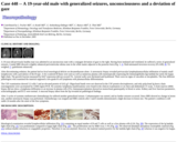
A 19-year-old previously healthy man was admitted in an unconscious state with a conjugate deviation of gaze to the right. Having been intubated and ventilated, he suffered a series of generalized seizures. Cranial MRI showed a slightly enhanced periventricular edema zone in the white matter adjacent to the posterior horn (Fig. 1; A: fluid-attenuated inversion recovery [FLAIR], B: T2-weighed, C: gadolinium enhanced).
(This case study was added to OER Commons as one of a …
(This case study was added to OER Commons as one of a batch of over 700. It has relevant information which may include medical imagery, lab results, and history where relevant. A link to the final diagnosis can be found at the end of the case study for review. The first paragraph of the case study -- typically, but not always the clinical presentation -- is provided below.)
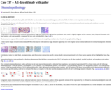
A 1-day old male was found to have pallor after birth. He was the product of an uneventful pregnancy and normal birth. He had no overt congenital anomalies/stigmata.
No restrictions on your remixing, redistributing, or making derivative works. Give credit to the author, as required.
Your remixing, redistributing, or making derivatives works comes with some restrictions, including how it is shared.
Your redistributing comes with some restrictions. Do not remix or make derivative works.
Most restrictive license type. Prohibits most uses, sharing, and any changes.
Copyrighted materials, available under Fair Use and the TEACH Act for US-based educators, or other custom arrangements. Go to the resource provider to see their individual restrictions.