(This case study was added to OER Commons as one of a …
(This case study was added to OER Commons as one of a batch of over 700. It has relevant information which may include medical imagery, lab results, and history where relevant. A link to the final diagnosis can be found at the end of the case study for review. The first paragraph of the case study -- typically, but not always the clinical presentation -- is provided below.)
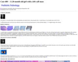
The patient is a 20 month old girl with an unremarkable past medical history. She presented with a 1 month history of a non-tender mass in her left calf.
An MRI demonstrated a soft tissue mass within the muscles of the left leg located posterior to tibia and fibula (Figure 1; T1 weighted image with contrast).
(This case study was added to OER Commons as one of a …
(This case study was added to OER Commons as one of a batch of over 700. It has relevant information which may include medical imagery, lab results, and history where relevant. A link to the final diagnosis can be found at the end of the case study for review. The first paragraph of the case study -- typically, but not always the clinical presentation -- is provided below.)
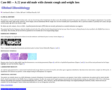
A 20 year old male with persistent cough was evaluated for surgical and anesthesia risk. The patient did not report fever, weight loss or night sweats. He was a smoker but quit 5yrs ago. No history of exposures to asbestos or industrial dusts was elicited.
(This case study was added to OER Commons as one of a …
(This case study was added to OER Commons as one of a batch of over 700. It has relevant information which may include medical imagery, lab results, and history where relevant. A link to the final diagnosis can be found at the end of the case study for review. The first paragraph of the case study -- typically, but not always the clinical presentation -- is provided below.)
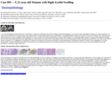
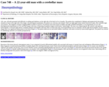
A previously healthy 20-year-old man developed progressive hearing loss for the last 3 years, evolving to tetraparesis with sphincter impairment. There is no history of fever, headache or vision loss. Neurologic examination demonstrated global spastic tetraparesis, more severe in lower limbs, with evident pyramidal signs (tetrahyperreflexia, clonus and bilateral Babinski sign) and severe bilateral hearing loss. Evaluation of sensibility and coordination was not reliable due to motor deficits and hearing loss.
(This case study was added to OER Commons as one of a …
(This case study was added to OER Commons as one of a batch of over 700. It has relevant information which may include medical imagery, lab results, and history where relevant. A link to the final diagnosis can be found at the end of the case study for review. The first paragraph of the case study -- typically, but not always the clinical presentation -- is provided below.)
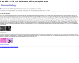
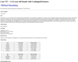
A 20 year old woman attended her general practitioner with right upper limb pain and intermittent paraesthesias for a 4 month period. She had no neck pain or systemic symptoms and was a non smoker. There was no family history of note. She was commenced on pregabalin for pain and an MRI of cervical spine was ordered. The MRI revealed a right-sided intradural extramedullary mass extending from C7-T1 that was displacing the spinal cord to the left. She was reviewed by neurosurgery and was now complaining of paraesthesias in the right lower limb also. She had no bowel or bladder symptoms. Her examination revealed reduced sensation in the right upper limb but normal tone, power, coordination and reflexes. Cranial nerves, the left upper limb and bilateral lower limb examination were documented as normal. Imaging revealed that the lesion now extended from C5 to T3 and was causing significant cord compression at C7-T1. An enhancing extradural soft tissue mass centered in and expanding the exit foramina and indenting the thecal sac (arrow) is shown by axial T1 MRI post-contrast (Figure 1). On the T2 Sagittal MRI (Figure 2) the low signal soft tissue mass is demonstrated in the right exit foramina at four spinal levels(small arrows), normal high signal fat is seen in the foramen below (larger arrow). (Figure 2) The patient underwent emergency resection of the lesion. A large rubbery, tan piece of tissue measuring 1.5 x 0.8 x 0.5 cms and further multiple pieces of cream grey tissue measuring 3.5 x 3 x up to 0.3 cm in aggregate were removed.
(This case study was added to OER Commons as one of a …
(This case study was added to OER Commons as one of a batch of over 700. It has relevant information which may include medical imagery, lab results, and history where relevant. A link to the final diagnosis can be found at the end of the case study for review. The first paragraph of the case study -- typically, but not always the clinical presentation -- is provided below.)
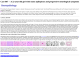
A previously healthy 21-month-old male presented with a 1-month history of vomiting, gait disturbance, and abnormal ocular alignment. He had papilledema, but no convulsions or abnormal consciousness. A CT scan revealed hydrocephalus (Figure 1a) and a cerebellar tumor measuring 4 cm in diameter (Figure 1b). Apparent diffusion coefficient map demonstrated strong restricted diffusion in the tumor (Figure 1c). The tumor was totally resected on the day after admission, and had not spread into his cerebrospinal fluid at surgery; however, the tumor progression was very aggressive. Only two weeks after surgery, and prior to any chemotherapy, the tumor had spread to his spinal cord leptomeninges, causing neoplastic meningitis. Moreover, although he was in remission after a cycle of chemotherapy (cyclophosphamide, cisplatin, vincristine, etoposide, and intrathecal methotrexate), the tumor relapsed quickly during additional chemotherapy. We prescribed an anthracycline-containing regimen and irradiation sequentially; however, the tumor did not regress. He survived only seven months after his diagnosis.
(This case study was added to OER Commons as one of a …
(This case study was added to OER Commons as one of a batch of over 700. It has relevant information which may include medical imagery, lab results, and history where relevant. A link to the final diagnosis can be found at the end of the case study for review. The first paragraph of the case study -- typically, but not always the clinical presentation -- is provided below.)
A 21 month old female child of non consanguineous parentage was admitted to the neurosurgical unit with rapidly progressing spastic paraparesis of lower limbs, following a febrile episode of 20 days duration. The developmental milestones were normal, with no history of trauma or any significant illness or hospitalization in the past. MRI of the dorsal spine revealed a large T1 - T4 right paravertebral circumscribed mass with intraspinal extension and severe cord compression suggestive of a neurofibroma with dumb-bell shaped intra and extraspinal extension (Fig. 1 and 2). On ultrasound examination there was no evidence of an abdominal mass. The tumor was approached through a right posterolateral thoracotomy. A greenish pink tumor adherent to the parietal pleura and right T2 - T4 intercostal nerves, vagus nerve and extending as small lobules into the tracheo- esophageal groove and carina was excised, leaving a small portion adherent to the vessels. Resecting the posterior ends of second, third and fourth ribs on the right and nibbling the vertebral pedicles from T1 - T4, the intraspinal portion of the lesion was totally resected. Post operatively the child had chylothorax that regressed spontaneously after 4 weeks. Chemotherapy was advised, but the parents refused. On follow up after one year, the child had regained normal power of lower limbs, but had mild spasticity. A repeat MRI after one year revealed no increase in size of the residual lesion and no evidence of metastatic disease anywhere (Fig. 3).
(This case study was added to OER Commons as one of a …
(This case study was added to OER Commons as one of a batch of over 700. It has relevant information which may include medical imagery, lab results, and history where relevant. A link to the final diagnosis can be found at the end of the case study for review. The first paragraph of the case study -- typically, but not always the clinical presentation -- is provided below.)
A 21-year-old female with a history of elevated blood pressure as a child and right nephrectomy for pyelonephritis. In 2003, she underwent an excision of a bladder mass.
(This case study was added to OER Commons as one of a …
(This case study was added to OER Commons as one of a batch of over 700. It has relevant information which may include medical imagery, lab results, and history where relevant. A link to the final diagnosis can be found at the end of the case study for review. The first paragraph of the case study -- typically, but not always the clinical presentation -- is provided below.)
A 21-year-old female admitted to the hospital with severe headache. While driving, she suddenly could not feel the steering wheel in her right hand and experienced a feeling of getting lost. The symptoms subsided but followed by headache that gradually evolved into the worst headache in her life. Neurologic exam was normal and MRI imaging disclosed left parietal tumor with a large cystic component and enhancing mural nodule (Figure 1A). Differential diagnosis included ganglion cell tumor, PXA and pilocytic astrocytoma. The patient underwent left craniotomy with a complete resection of the tumor. Intraoperatively, the mass was intra-axial and consisted of a white-yellow firm 3 cm nodule located on a wall of a large cavity filled with an amber-colored fluid. There was no association with leptomeninges.
(This case study was added to OER Commons as one of a …
(This case study was added to OER Commons as one of a batch of over 700. It has relevant information which may include medical imagery, lab results, and history where relevant. A link to the final diagnosis can be found at the end of the case study for review. The first paragraph of the case study -- typically, but not always the clinical presentation -- is provided below.)
A previously healthy 21-year-old female presented with permanent tinnitus, vertigo and nausea lasting for two months. Physical examination did not reveal any neurological deficits. Her family history was negative. All blood tests, including serum levels of the germ cell tumor markers alpha-fetoprotein (1.1 µg/l) and beta-human chorion gonadotropin (< 2 mIU/ml), were normal. Magnetic resonance imaging (MRI) of the brain demonstrated a small, partially cystic, contrast-enhancing mass in the posterior part of the third ventricle. Fig. 1a shows the tumor (arrow) on a contrast-enhanced T1-weighted sagittal MRI scan. Fig. 1b demonstrates a coronal section through the tumor. The tumor did not result in an obstruction of the cerebrospinal fluid flow. No further intracranial lesions were present. We additionally performed a digital subtraction angiography, which revealed a variant of the vein of Galen but no pathological vascularization of the intraventricular mass. The tumor was resected via an infratentorial supracerebellar approach. The postoperative course was uneventful.
(This case study was added to OER Commons as one of a …
(This case study was added to OER Commons as one of a batch of over 700. It has relevant information which may include medical imagery, lab results, and history where relevant. A link to the final diagnosis can be found at the end of the case study for review. The first paragraph of the case study -- typically, but not always the clinical presentation -- is provided below.)
A 21-year-old female patient, without previous co-morbidities, presented with progressive decrease in visual acuity in the right eye, ipsilateral ptosis, frontal headache and bilateral galactorrhea on expression in the last seven months before the consultation. At that time, in another institution, a serum prolactin level, which was 74.9 ng/mL [normal range (NR): 5 - 25], and a sella turcica magnetic resonance imaging (MRI) were requested. The MRI (Figures 1, 2, 3 and 4) revealed an expansive solid lesion, isointense in T1- and T2-weighted sequences with gadolinium enhancement measuring 3.5x3.3x4.2 cm, with its epicenter in the sella turcica. It extends superiorly reaching the optic chiasm, laterally mainly to the right cavernous sinus and inferiorly towards the sellar floor, causing erosion of the sellar floor and the adjacent area of the clivus. She was treated with bromocriptine 2.5 mg/day for two months.
(This case study was added to OER Commons as one of a …
(This case study was added to OER Commons as one of a batch of over 700. It has relevant information which may include medical imagery, lab results, and history where relevant. A link to the final diagnosis can be found at the end of the case study for review. The first paragraph of the case study -- typically, but not always the clinical presentation -- is provided below.)
21-year-old non-diabetic man presented with acute renal failure, approximately 2 weeks following a "sore throat". The patient has a history of HIV and hepatitis. Pertinent laboratory investigations: Creatinine: 4.5 mg/dl, BUN: 65 mg/dl, potassium: 6.0 mEq/ml, ANA: not available, anti-double stranded DNA antibody: negative, ANCA: negative, anti-GBM antibody: negative, ASO antibody: negative, C3: 98 mg/dl, C4: 28 mg/dl, HIV antibody: positive, hepatitis B surface antibody: positive, hepatitis B surface antigen: negative, hepatitis C antibody: negative, rheumatoid factor: negative, SPEP / UPEP: no evidence of a monoclonal immunoglobulin. Urine sediment: contains red blood cells, urine protein: 2.5 gm/24 hrs, Urine toxicology: positive for marijuana and opiates.
(This case study was added to OER Commons as one of a …
(This case study was added to OER Commons as one of a batch of over 700. It has relevant information which may include medical imagery, lab results, and history where relevant. A link to the final diagnosis can be found at the end of the case study for review. The first paragraph of the case study -- typically, but not always the clinical presentation -- is provided below.)
A 21-year old male with a history of intravenous heroin abuse presented to Presbyterian Hospital status post cardiac arrest. The patient was found unconscious by his father with snoring respirations. His father turned to call 911. When he returned from that phone call, he found that his son had stopped breathing. He was found with Seroquel (quetiapine) packets in his room. When paramedics arrived the patient was in asystole.
(This case study was added to OER Commons as one of a …
(This case study was added to OER Commons as one of a batch of over 700. It has relevant information which may include medical imagery, lab results, and history where relevant. A link to the final diagnosis can be found at the end of the case study for review. The first paragraph of the case study -- typically, but not always the clinical presentation -- is provided below.)
This 21 year-old male had a history of posterior urethral valves and subsequent obstructive uropathy, requiring a single renal transplantation in 1989. He developed chronic allograft nephropathy in 2001 and underwent a second renal transplantation in 2006, with subsequent multiple episodes of rejection. His treatment included Cellcept, Prograf, methylprednisolone, intravenous immunoglobulin and plasmapheresis. He presented to the emergency room complaining of severe headaches, nuchal rigidity, tachycardia and rigors.
(This case study was added to OER Commons as one of a …
(This case study was added to OER Commons as one of a batch of over 700. It has relevant information which may include medical imagery, lab results, and history where relevant. A link to the final diagnosis can be found at the end of the case study for review. The first paragraph of the case study -- typically, but not always the clinical presentation -- is provided below.)
A 22-year old female presented with a five-week history of right retroorbital pain and eyelid swelling. She denied headaches, loss of vision, focal neurological symptoms or ocular symptoms. On exam she had tenderness over right eyebrow and temporal bone. Her past medical history was unremarkable. An ophthalmologist initially saw her and a CT-scan was ordered. The scan showed an orbital bony lesion, which contained a solid component as well as a fluid level without calcifications or a bony matrix (Figure 1). A subsequent MRI revealed a contrast-enhancing lesion eroding the roof of the orbit posterolaterally on the right (Figure 2). Involvement of the inner table of the temporal bone was also demonstrated on MRI. The patient was referred to our center at this time for further evaluation. A CT angiogram was performed to assess the vascularity of the lesion and whether or not any preoperative embolization would be of utility. On the angiogram no vascular abnormalities could be identified intracranially. One month after her initial presentation she successfully underwent a bifrontal craniotomy with resection of the orbito-temporal lesion and placement of a prosthetic right orbital roof.
(This case study was added to OER Commons as one of a …
(This case study was added to OER Commons as one of a batch of over 700. It has relevant information which may include medical imagery, lab results, and history where relevant. A link to the final diagnosis can be found at the end of the case study for review. The first paragraph of the case study -- typically, but not always the clinical presentation -- is provided below.)
A 22-year-old girl presented with convulsive status epilepticus and a previous history of recurrent seizures, myoclonus, ataxia and impaired cognitive functions. Testing revealed severe metabolic acidosis, elevated transaminases and creatine kinase, and respiratory insufficiency. After intubation and ventilation, thiopental was introduced but the patient's condition worsened dramatically with death after a few hours. The parents gave permission for autopsy.
(This case study was added to OER Commons as one of a …
(This case study was added to OER Commons as one of a batch of over 700. It has relevant information which may include medical imagery, lab results, and history where relevant. A link to the final diagnosis can be found at the end of the case study for review. The first paragraph of the case study -- typically, but not always the clinical presentation -- is provided below.)
The patient is a 22 year old Saudi Arabian male, who has been living in the United States for three years. He initially presented 2 days prior to admission to the emergency department of an outside hospital with cough, night sweats, anorexia, severe fatigue, and an unintentional 90 lbs weight loss over the 6 months prior to presentation. He is a previous smoker (1 pack/day for 8 years), but quit several months ago due to his chronic cough.
(This case study was added to OER Commons as one of a …
(This case study was added to OER Commons as one of a batch of over 700. It has relevant information which may include medical imagery, lab results, and history where relevant. A link to the final diagnosis can be found at the end of the case study for review. The first paragraph of the case study -- typically, but not always the clinical presentation -- is provided below.)
A26- year- old male presented with difficulty in walking and shakiness on the right side of the body for 8 to 9 months. The patient also complained of diplopia and progressively decreasing sensation of taste on the right side of the tongue. On neurological examination, the patient was conscious and oriented. There was right upper motor neuron facial nerve palsy. The cerebellar signs were positive. The magnetic resonance imaging (MRI) revealed 4.7x4.6x4.3 cm posterior fossa mass lesion in midline and right cerebellar hemisphere. It was hypointense on T1w and heterogeneously hyperintense on T2w and flair images with areas of blooming on gradient images (calcification / hemorrhage). The mass was compressing the 4th ventricle with upstream hydrocephalus. It involved right inferior cerebellar peduncle and medulla with mass effect on brain stem and a focal hyperintense focus in continuity with the right dorsal medulla. On post-contrast images there was significant enhancement of the lesion (Figure 1). The patient underwent an uneventful midline craniotomy with tumor excision by transvermian approach. The patient was referred for radiotherapy and alive at 6 months postoperatively.
(This case study was added to OER Commons as one of a …
(This case study was added to OER Commons as one of a batch of over 700. It has relevant information which may include medical imagery, lab results, and history where relevant. A link to the final diagnosis can be found at the end of the case study for review. The first paragraph of the case study -- typically, but not always the clinical presentation -- is provided below.)
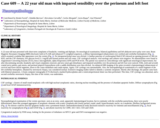
A 22-year-old man presented with short-term complaints of headache, vomiting and diplopia. On neurological examination, bilateral papilledema and left abducent nerve palsy were seen. Brain Magnetic Resonance Imaging (MRI) disclosed, both in FLAIR and enhanced T1-weighted sequences, a diffuse leptomeningeal enhancement over cerebral and cerebellar hemispheres (Fig. 1), which assumed a nodular-like appearance over the vermis (Fig. 2). No intraparenchymal mass was elicited. A lumbar puncture of cerebrospinal fluid (CSF) showed an opening pressure of 360 cm H2O, and the CSF chemistry, low glucose level (1.1 mg/dl) and normal protein and cell count. CSF cytological analysis, viral serology and cultures were negative. No abnormalities were found in Angiotensin-Converting Enzyme (ACE), beta-2 microglobulin, alpha-fetoprotein (AFP) and PCR levels. The patient was started on corticotherapy with significant neurological improvement, but after discontinuing steroids, headache and visual complaints renewed, and new-onset gait disturbance and impaired sensibility over the perineum and left foot were noticed. Fifth, sixth and seventh cranial nerve palsies, gait ataxia, and perineal pinprick hypoesthesia with a saddle distribution were then elicited. An enhanced MR imaging of the spine revealed a leptomeningeal enhancement of cervical and lower dorsal segments, down to the conus medullaris and cauda equine. Again, CSF cytology was unremarkable. Also, the neuropathological examination of a small fragment of the left cerebellar hemisphere, obtained by stereotactic biopsy, was considered normal. Extensive imaging studies failed to identify a systemic neoplasm. Shortly after discharge, the patient came back with bilateral amaurosis. A brain computer tomography (CT) disclosed a marked hydrocephalus and a ventriculoperitoneal shunt was then performed. This time, CSF cytology was abnormal, and a second cerebellar stereotactic biopsy, this time of the vermis, was undertaken.
(This case study was added to OER Commons as one of a …
(This case study was added to OER Commons as one of a batch of over 700. It has relevant information which may include medical imagery, lab results, and history where relevant. A link to the final diagnosis can be found at the end of the case study for review. The first paragraph of the case study -- typically, but not always the clinical presentation -- is provided below.)
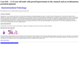
The patient is a 23 year old male with a history of Crohn's disease and primary sclerosing cholangitis since age 15. In 2003 he received a living donor liver, which had to be replaced with an orthotopic liver 10 days after transplant due to complications. Over the previous month, his liver enzymes had been trending upwards, worrisome for return of primary liver disease, versus possible stricture at the biliary-enteric anastomosis. Aside from mild generalized jaundice, no discrete worrisome skin lesions were noted. He complained of abdominal and back pain, the latter believed to be a mild inflammation of spinal joints. An upper endoscopy was performed, and significant findings included a patent anastomosis, evidence of portal hypertension in the stomach, and an erythematous proximal jejunum, which was biopsied. Of note, his pre-operative labs showed a microcytic anemia (hemoglobin 10.5 g/dL and MCV 73.9 fL) with an expanded red cell width of 19.5%, which was being treated with oral iron supplementation.
No restrictions on your remixing, redistributing, or making derivative works. Give credit to the author, as required.
Your remixing, redistributing, or making derivatives works comes with some restrictions, including how it is shared.
Your redistributing comes with some restrictions. Do not remix or make derivative works.
Most restrictive license type. Prohibits most uses, sharing, and any changes.
Copyrighted materials, available under Fair Use and the TEACH Act for US-based educators, or other custom arrangements. Go to the resource provider to see their individual restrictions.