(This case study was added to OER Commons as one of a batch of over 700. It has relevant information which may include medical imagery, lab results, and history where relevant. A link to the final diagnosis can be found at the end of the case study for review. The first paragraph of the case study -- typically, but not always the clinical presentation -- is provided below.)
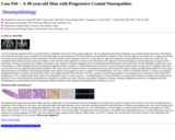
A 49-year-old man presented with a two-month history of headache and recent twenty pound weight loss. He was diagnosed with cluster headaches at an outside hospital and treated with triptans. One week after the initial onset of headache, he developed right jaw and facial pain with facial weakness, accompanied by inability to fully close his right eye and mouth. He was given one dosage of steroids at an outside hospital for cluster headache with full resolution of his symptoms. However, a week later, his right facial weakness recurred and persisted. About four weeks later, he complained about similar jaw/facial pain and weakness of his left side, which appeared to be more severe than those of his right side. He also developed occasional diplopia on far gaze and left lateral gaze. He did not experience any changes in hearing or taste. Magnetic resonance imaging of brain revealed abnormal enhancement of the third, fifth, seventh and eighth cranial nerves with various degree of diffuse enlargement (arrows pointing to trigeminal nerves in Figure 1). Patchy enhancement of the nerve roots in the distal lumbar region was also noted. A gallium whole body scan did not reveal any significant abnormality. Repeated CSF studies showed low glucose levels, elevated protein levels and elevated cell counts. Extensive peripheral blood and CSF work-up for infectious disease, autoimmune, demyelinating disorders and neoplasia were negative except for a positive T-cell lymphotropic virus type 1 (HTLV-1) antibody. His HIV-1/2 ELISA was negative. Peripheral blood flow cytometry showed mature WBCs with slightly elevated CD4:CD8 ratio. CSF flow cytometry revealed large granular T-cell lymphocytosis consisting of CD3/CD56/CD57+ lymphocytes (>97% of gated lymphocytes), which was interpreted as due to a reactive process. A working diagnosis of neurosarcoidosis was made and the patient was treated with prednisone 60 mg daily for two weeks followed by dexamethasone 4 mg daily for another two weeks. However, the patient's symptoms worsened, leading to heightened suspicion of a malignant neoplasm. Dexamethasone was discontinued and he subsequently underwent biopsy of the enlarged left infraorbital nerve (Figure 2, arrows).
- Subject:
- Applied Science
- Education
- Health, Medicine and Nursing
- Life Science
- Material Type:
- Case Study
- Diagram/Illustration
- Provider:
- University of Pittsburgh School of Medicine
- Provider Set:
- Department of Pathology
- Author:
- Charles Shao
- Constantine A. Axiotis
- Jenny Libien
- Jiancong Liang
- Jinli Liu
- Yuetsu Tanaka
- Date Added:
- 08/01/2022
